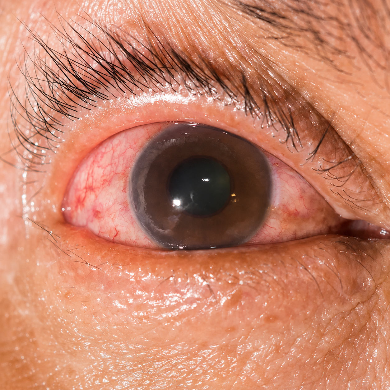
The retina is the light-sensitive layer at the back of your eye, and it relies on tiny veins to carry blood away from it. Retinal vein occlusion happens when one of those veins becomes blocked — usually by a blood clot, or by narrowing linked to conditions like high blood pressure or diabetes. When the vein is blocked, blood and fluid back up into the retina. That leakage causes macular edema (swelling of the macula, the central part of the retina you read and recognise faces with), which is the main reason vision drops in RVO.
RVO is not rare. It is the second most common retinal vascular disorder after diabetic eye disease, and it becomes more common with age. It is closely tied to blood-pressure, blood-sugar and cholesterol control — which is why treating the eye and reviewing your general health go hand in hand.
Because the vision loss is usually painless and in one eye, many people notice it only when they happen to cover the other eye. If your sight has changed suddenly, our retina treatment team can assess you the same week — early review gives the eye its best chance.
people worldwide are affected by retinal vein occlusion — the second most common retinal vascular disorder after diabetic eye disease.
Rogers et al., pooled analysis, Ophthalmology, 2010.
RVO is grouped by where the blockage sits and how much of the retina it starves of blood flow. Which type you have shapes the treatment plan and what to expect.
of vein occlusions are the branch form, according to international data — and Indian hospital data report a similar pattern.
Song et al., systematic review, Journal of Global Health, 2019.
A non-ischemic RVO can turn ischemic over time, so even a ‘mild’ occlusion is worth reviewing regularly. Scheduled scans tell us which way your eye is heading — long before you would notice the difference yourself.
RVO usually affects one eye and comes on without pain. See a retina specialist promptly if you notice any of these — which of them sounds like you?
Sudden blurring or a drop in vision in one eye, often noticed on waking.
A dark, grey or missing patch in part of your field of vision.
Distorted vision — straight lines look bent or wavy (a sign of macular swelling).
New floaters, or a sudden shower of spots (can signal bleeding inside the eye).
Vision that fluctuates — clearer at some times, blurrier at others.
Sudden vision loss should always be checked quickly — book a comprehensive eye examination rather than waiting to see if it settles.
Pain and redness are not typical early on. If they appear later, they can point to raised eye pressure and need urgent attention. A sudden drop in vision in one eye should be examined the same day wherever possible.
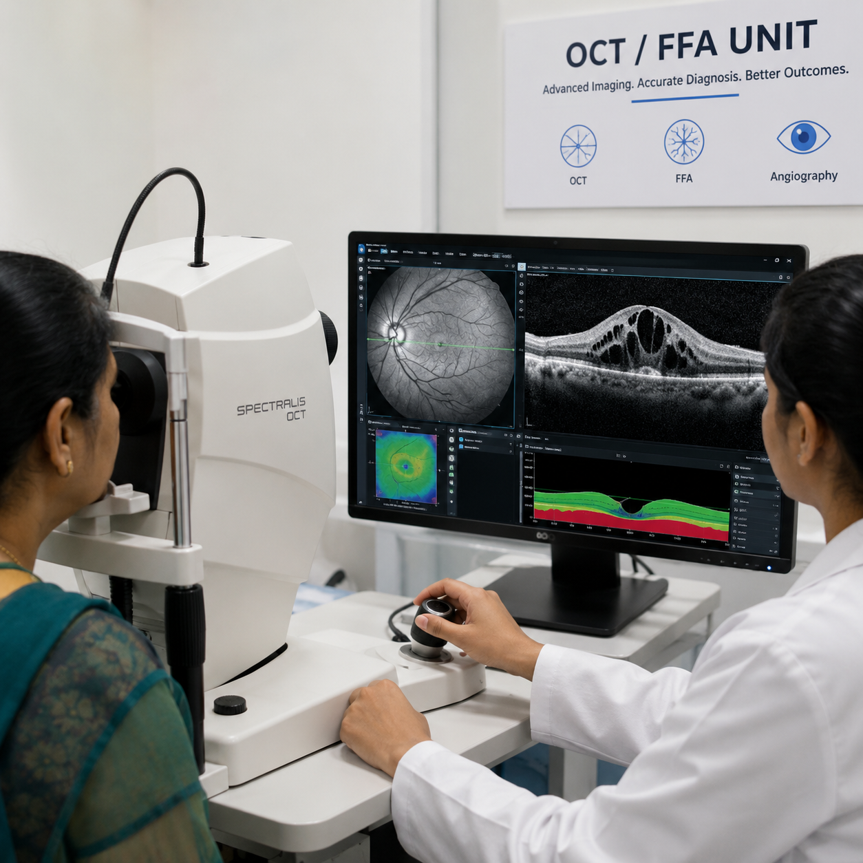
Call +91 81786 39701RVO is often recognisable the moment a retina specialist examines the back of your eye, but confirming the type and measuring the swelling guides the whole treatment plan.
These scans are repeated over the treatment course so we can see, objectively, whether the swelling is settling — not guess. Distinguishing RVO from look-alike conditions such as diabetic retinopathy is part of this assessment.
To see the pattern of retinal haemorrhages and confirm whether the occlusion is branch or central.
A quick, non-invasive scan that maps and measures macular swelling and tracks your response to treatment.
A dye study that shows which vessels are leaking or closed, and whether the occlusion is ischemic.
Including blood pressure, blood sugar and cholesterol, because RVO is often the eye’s warning sign of a wider vascular issue.
The goal of treatment is to control the macular swelling that reduces vision and to protect the eye from complications. RVO is managed, not switched off in a single step — but with timely, structured treatment many patients regain useful vision. Our retina team tailors the plan to your type of RVO, how much swelling is present and your general health.
Anti-VEGF medicines (such as ranibizumab, aflibercept and bevacizumab) are injected into the eye to reduce leakage and swelling. These are treatment averages, not a promise for any one eye — your outcome depends on the type and severity of your RVO and how early treatment starts.
Injections are given after numbing drops, so most patients feel pressure rather than sharp pain. RVO usually needs a series of injections over months — sometimes longer — with OCT scans guiding how many. Anti-VEGF reduces swelling but does not ‘cure’ the blocked vein, which is why regular follow-up matters (American Society of Retina Specialists).
of branch-RVO patients gained at least three lines of vision at six months with monthly ranibizumab, versus about 29% on a sham injection.
of central-RVO patients gained three or more lines, versus roughly 17% on sham.
BRAVO and CRUISE trials, Ophthalmology, 2010.
Where the retina is ischemic and starts to grow abnormal, fragile vessels, targeted laser (including pan-retinal photocoagulation) helps calm that response and lowers the risk of bleeding and a difficult form of glaucoma. Laser may be used alongside injections rather than instead of them.
For selected patients, a steroid injection or a slow-release steroid implant (such as dexamethasone) is an alternative way to control stubborn swelling. Because steroids can raise eye pressure or speed cataract formation, we choose them case by case and monitor closely.
Treating underlying causes is part of the plan too — good control of blood pressure, diabetes and cholesterol supports the eye and lowers the chance of the other eye being affected. Where nutrition plays a supporting role in retinal health, we may also discuss eye nutrition treatment. Not every eye responds the same way; if a different retinal condition is contributing, we will guide you to the right treatment.
Consult with our doctorsIndividual results vary. Suitability, benefits and risks depend on the type and severity of your occlusion, how much swelling is present and your overall health, and can only be determined after a detailed examination with our doctors.
You cannot always prevent RVO, but you can lower your risk and protect your second eye by managing the vascular factors behind it:
Keep blood pressure well controlled — hypertension is the single biggest modifiable risk factor for RVO.
Manage diabetes and keep blood sugar in your target range.
Control cholesterol and follow a heart-healthy routine — regular activity, no smoking.
Have regular eye checks if you have glaucoma or a family history of eye disease.
Report any sudden change in vision straight away — early treatment protects sight.
RVO is often the eye’s early signal of a wider circulation problem, so treating the eye is only half the job. We work alongside your physician to review blood pressure, blood sugar and cholesterol, and we recommend a general medical check after diagnosis — especially if both eyes are involved, where clotting tendencies are more likely and further tests may be advised (American Society of Retina Specialists).
Managing these conditions well supports your treated eye and helps safeguard the other. Our retina specialists also monitor for related retinal disease such as Diabetic Retinopathy and Age-related Macular Degeneration (AMD), which can coexist and affect the plan.
Cost depends on the type of RVO, how much swelling is present, the medicine chosen and how many injections your eye needs over the treatment course. The figures below are indicative starting-from ranges to help you plan — they are not a fixed quote, and your exact cost is confirmed only after a consultation and eye examination.
Indicative ranges from Dayal Eye Centre’s retina fee context (verified clinical data 2024–2025); subject to re-verification and to your individual consultation. Individual results and costs vary based on eye condition, severity, medicine used, number of injections and healing response.
Easy EMI options (via Savein) to spread the cost of treatment.
Cashless insurance / TPA support where your policy covers retinal treatment.
CGHS / ECHS / Ayushman Bharat / Haryana Government scheme assistance where eligible.
Want a clear estimate for your eye? We’ll explain what’s included before anything begins.
Consult with our doctorsRVO rewards early, specialist-led care with the right diagnostics on hand. At Dayal Eye Centre you get that in one place:
Fellowship-trained retina team — dedicated retinal specialists who manage RVO, diabetic eye disease and macular conditions day in, day out.
Diagnostics under one roof — OCT and fluorescein angiography on site, so diagnosis and monitoring don’t mean multiple trips.
Full treatment ladder — anti-VEGF injections, retinal laser and intravitreal steroids, matched to your type and severity of RVO.
NABH-accredited, 20+ years, 1.2 lakh+ surgeries — an accredited single centre with a senior team and a strong safety culture.
Co-ordinated, whole-person care — we treat the eye and work with your physician on the blood-pressure and sugar control that protects it.
Consult with our doctors at Dayal Eye Centre — one centre, one senior retina team.
808 B, Galaxy Road, Sector 15 Part 2, Near 32nd Avenue, Gurgaon, Haryana 122001
+91 81786 39701
wecare@dayaleyecentre.in
Mon–Sat, 8:30 AM – 7:30 PM · Sunday closed. Beside 32nd Avenue, off NH-48 · ~25 min from Delhi · nearest metro IFFCO Chowk (Yellow Line).
The questions patients ask us most about retinal vein occlusion. For anything specific to your eye, a consultation gives the clearest answer.
Medically reviewed by Dr. Neelam Khatwani and the retina team at Dayal Eye Centre · Last reviewed: 29 July 2026 · Editorial Policy