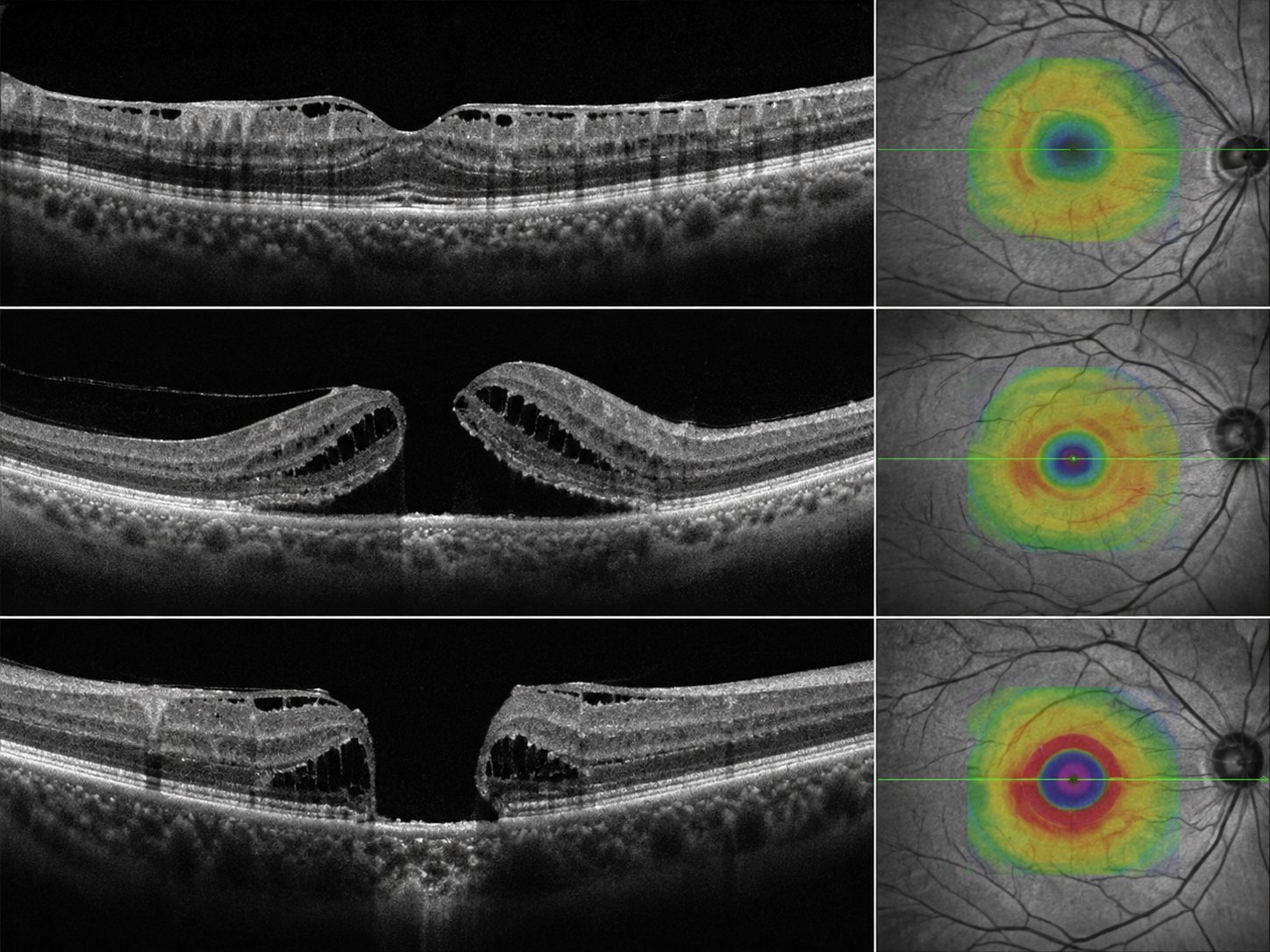
The macula is the part of the retina responsible for your sharp central vision, and ERM, VMT and macular hole all interfere with how it works — causing distortion, blur or a blind spot in the middle of your sight.
Early diagnosis and the right treatment can protect that central vision and, in many cases, improve it. Learn more about our full Retina Treatment options in Gurgaon.
ERM, VMT and macular hole are related conditions of the macula that share overlapping symptoms. Telling them apart on an OCT scan is what decides whether you need watching or treatment.
Also called a macular pucker
An epiretinal membrane is a thin sheet of scar-like tissue that forms on the surface of the retina, usually over the macula. As it contracts it wrinkles or “puckers” the retina beneath — which is why it is often called a macular pucker — and this is what makes straight lines look bent and central vision blurry.
ERM is most often linked to normal age-related changes in the vitreous gel, but it can also follow a retinal tear, inflammation inside the eye, diabetic eye disease, or previous eye surgery. It is common and becomes more likely with age, and more common in eyes that have had cataract surgery. Large OCT-based studies (Beaver Dam Eye Study) have detected some degree of ERM in up to about a third of older adults — though most are mild and never affect sight.
The vitreous pulling on the macula
Vitreomacular traction happens when the vitreous — the clear gel that fills the eye — stays partly stuck to the macula as it naturally separates with age, and pulls on it. That tension stretches the macula and can cause blurred or distorted central vision, sometimes with flickering or a change in image size.
VMT can occur on its own with ageing, or alongside other retinal conditions such as diabetic retinopathy or macular degeneration. Some cases release by themselves and only need monitoring; others progress and benefit from treatment — which is exactly what an OCT scan and a retina consult help us tell apart.
A small break in the centre of the macula
A macular hole is a small, full-thickness break in the centre of the macula. Because it sits exactly where your sharpest vision is focused, it typically causes a dark or missing patch in the middle of your sight, along with distortion. Most come from age-related shrinking of the vitreous gel, but they can also follow an eye injury or occur in very short-sighted (highly myopic) eyes.
Idiopathic (no obvious cause) macular holes involve the second eye in around 1 in 9 patients — which is why we examine both eyes carefully. Unlike many ERMs, a macular hole rarely closes on its own and is usually treated with surgery to give the best chance of restoring central vision.
A membrane on a scan is not automatically a membrane that needs surgery. What counts is how much it is affecting your vision — which is exactly what an OCT scan and a retina consult help us tell apart. An epiretinal membrane forms on the surface of the retina, usually over the macula.
These conditions share overlapping symptoms. See how many of these sound like your day-to-day vision:
Near or distance vision loses its sharpness in the centre while the edges stay clear.
Door frames, tiles and text look distorted (metamorphopsia) — an important warning sign.
Difficulty reading, using a phone, or recognising faces as the macula is affected.
A blank spot in the middle of your vision — more typical of a macular hole.
Symptoms usually build up gradually and may be mild early on. But a sudden increase in floaters, flashes of light, or a curtain or shadow over your vision is different — it can signal a retinal tear or detachment and needs same-day attention.
Call +91 81786 39701A simple home check: look at each eye separately. If straight lines look distorted or a patch is missing, book an evaluation with our retina specialist Dr. Neelam Khatwani.
Diagnosing a macular condition takes more than a standard eye test. We combine a detailed examination with high-resolution imaging so we can see the macula in cross-section and measure exactly what is happening:
A direct look at the retina and macula to spot puckering, traction or a hole.
A quick, non-contact scan that produces detailed cross-section images of the macula. OCT is the key test for ERM, VMT and macular hole: it confirms the diagnosis, grades the severity, and guides whether surgery is needed.
A dye-based scan of the retinal blood vessels, used when we need to check for related problems such as diabetic damage or swelling.
Because OCT shows the macula layer by layer, it lets us track a mild membrane over time and act only when treatment will genuinely help — sparing you surgery you do not yet need.
Examination, OCT and angiography where needed — all in-house, so your plan is decided the same day.
Book an evaluationNot every macular condition needs an operation. The decision is between watching a mild, stable membrane and operating when distortion starts to interfere with daily life.
Many epiretinal membranes and some cases of VMT are mild and do not need immediate surgery — they can be safely watched with periodic OCT scans.
Simple aids such as good lighting and magnifiers, and keeping the prescription up to date, help manage minor distortion while we monitor for any change.
When distortion or blurring starts to interfere with daily life, the treatment is a vitrectomy — keyhole surgery through tiny ports, in which the surgeon removes the vitreous gel and addresses the problem at the macula. How this is done depends on which condition you have.
The surgeon gently peels the membrane (and often the fine inner limiting membrane) off the macula to release the pucker and let the retina flatten.
The surgery relieves the gel’s pull on the macula so it can settle back toward its normal shape.
The surgeon removes the vitreous and places a gas bubble that presses on the hole to help it close; a period of face-down positioning is usually advised afterwards.
These figures are from published medical research, not a promise of any individual result — the right expectation for your eye is something our surgeons set after examining it. Outcomes depend on how long the problem has been present and its severity, which is why an early, accurate assessment matters. Consult with our doctors to discuss whether surgery will help you.
Cost depends on which procedure you need, the anaesthesia, whether a gas or oil tamponade is used, and any imaging or follow-up care. Monitoring a mild membrane costs little beyond periodic scans; surgery is a bigger investment. As a guide only, and never a fixed quote:
These are starting-from ranges, subject to a consultation and eye examination — your final cost is confirmed only after we assess your eye.
We help make treatment manageable with Easy EMI (via Savein), cashless insurance and TPA support, and government schemes where applicable. Insurance coverage varies by plan — our team will help you check yours.
Prices are indicative starting ranges, subject to change; final cost is determined after a clinical consultation and depends on individual factors.
Vitrectomy is usually a day-care procedure, and vision recovers gradually. Here is what the weeks after surgery typically look like.
Vitrectomy is usually day-care. Some scratchiness, redness or mild discomfort in the first few days is normal and settles with prescribed drops.
For ERM and VMT, sharpness and distortion typically keep improving over several weeks to a few months.
Vision can continue to refine for up to a year as the macula relaxes. Distortion often eases over this period.
Regular OCT confirms the macula is settling, the hole has closed or the membrane has not returned, and catches any complication early.
If a gas bubble was used (mainly for macular holes), you may be asked to keep a face-down or specific head position for a set period, and you must not fly or travel to high altitude until the bubble has absorbed. Your surgeon will confirm when it is safe.
It is common to develop a cataract in the year or two after vitrectomy. This is expected, easily treated later, and something we watch for at your reviews with regular OCT to confirm the macula is settling.
No lifestyle step can guarantee you will never develop a macular condition, but these habits genuinely reduce risk and keep your eyes monitored.
Regular checks — especially after 50 or after cataract surgery — catch macular changes early, when monitoring or timely surgery gives the best outcome.
Keeping diabetes, blood pressure and high myopia well controlled lowers the risk of macular complications and helps existing conditions stay stable.
UV-protective sunglasses, a diet rich in omega-3 and antioxidants, and not smoking all support long-term retinal health.
Macular surgery is precise, specialist work, and where you have it done matters. You are cared for by a dedicated retina team inside an NABH-accredited centre with three modular operation theatres and an in-patient ward — all under one roof in Sector 15, Gurgaon.
Fellowship-trained retina specialists using OCT-guided diagnosis and small-gauge vitrectomy.
NABH-accredited facility · three modular OTs · in-patient ward — one centre, no run-around.
20+ years of experience · 1.2 lakh+ surgeries · Ayushman Bharat Awards 2021 & 2024.
Transparent, consultation-based pricing with EMI, cashless insurance and government schemes.
MBBS, DNB (Gold Medalist)
A fellowship-trained vitreo-retinal surgeon with 8+ years of experience, she leads medical and surgical retina care at Dayal Eye Centre. She treats ERM, VMT, macular hole and the wider spectrum of retinal disease, and personally sets realistic expectations before recommending surgery.
Read full profileThe questions patients ask us most about ERM, VMT and macular hole surgery. For anything specific to your eyes, a consultation gives the clearest answer.
Individual results depend on your specific condition, how long it has been present, age, eye health and healing. Statistics quoted are from published research, not a promise of any individual outcome.
Not every macular condition needs surgery, and not everyone is a candidate; suitability is decided only after a detailed examination.
All surgery carries some risk. Your surgeon will discuss the specific risks and benefits for your eye at consultation.
Prices are indicative starting ranges, subject to consultation and examination; insurance coverage varies by plan.
This page is for information and does not replace a consultation. Please consult a qualified eye specialist for diagnosis and treatment specific to your condition.
Medically reviewed by Dr. Neelam Khatwani, Vitreo-Retinal Surgeon, Dayal Eye Centre · Last reviewed: 29 July 2026 · Editorial Policy