Leave your number. A specialist calls you back today.
Copy the number, or leave your details and we will call you back.
Mon–Sat · 8:30 AM–7:30 PM · Sector 15, Gurugram
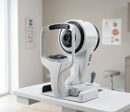
A rotating Scheimpflug camera that builds a 3D model of your cornea in seconds — the scan that tells us whether LASIK, SMILE or Contoura Vision is a safe choice for your eyes.
Before we reshape a cornea with a laser, we need to know it in three dimensions — how thick it is at every point, how it curves, and whether its shape is behaving normally. The OCULUS Pentacam is the scan that tells us, in a few seconds per eye, with nothing touching your eye. It often decides whether LASIK, SMILE or Contoura Vision is a safe choice for you.
It sits behind every laser vision correction workup we do in Gurgaon, behind keratoconus screening and behind premium-lens cataract planning — a diagnostic and planning instrument that does not treat anything.
It is a rotating Scheimpflug camera: it sweeps a slit of light around your eye and rebuilds photographs taken from many angles into a three-dimensional model — tomography, meaning depth rather than a flat picture of the surface. In one scan it captures:
The cornea is the clear front window of your eye. Older machines mapped only its front; the Pentacam also maps the back, where the earliest signs of weakening appear first.
Pachymetry means corneal thickness, measured across the whole cornea rather than at one point, so we see the thinnest spot and where it sits — the figure that limits how much tissue a laser can safely remove.
How steeply the cornea domes, and how far each point rises above or falls below the shape a normal cornea should have — revealing astigmatism and any irregular bulge.
The anterior chamber is the fluid-filled space between the cornea and the coloured iris. Its depth matters for implantable lenses; its drainage angle bears on glaucoma (kala motia) risk.
How cloudy your natural lens has become — helping decide whether blurring is a cataract (motiyabind) rather than your glasses number.
Laser correction removes a very thin layer of corneal tissue to change your focus. That is only sensible if your cornea is thick enough to give tissue away and stay strong, and regular enough to behave predictably afterwards.
The Pentacam answers both. If the map shows a cornea too thin for the correction your power needs, flap-based LASIK may be off the table and a surface procedure such as PRK discussed instead. If the elevation maps show an irregular or suspicious pattern, laser correction may not be advisable at all.
Saying no when the map says no is the point of doing this test. Suitability depends on a detailed evaluation — not every patient is a candidate, and we will tell you plainly if you are not.
Where the scan is reassuring, it shapes which procedure we recommend — including SMILE and topography-guided Contoura Vision — and feeds into how it is planned.
Keratoconus is a condition in which the cornea gradually thins and bulges into a cone shape, blurring and distorting vision. Early on it causes nothing you would notice — just a glasses power that keeps changing.
Because the Pentacam reads the back surface and thickness across the whole cornea, it picks up these patterns before a routine eye test would. Operating on a cornea that is already quietly weakening is what screening before laser surgery exists to prevent.
The same scans then track the condition. Comparing maps over time tells us whether keratoconus is stable or progressing, and progression is the usual trigger for corneal collagen cross-linking (CXL), which strengthens corneal tissue to slow further change. Pentacam readings also guide whether CXL can be done safely and how it is planned. More on our Keratoconus page.
The power of the artificial lens (IOL) replacing your natural lens comes chiefly from optical biometry on the ZEISS IOLMaster 700. Pentacam data is used alongside it for toric and premium lenses, where true corneal astigmatism and anterior chamber depth affect the result.
Anterior chamber depth and drainage angle help identify eyes at risk of narrow-angle glaucoma. Corneal thickness also changes how eye pressure readings are interpreted — a thick cornea reads higher than the truth, a thin one lower.
After laser correction, cross-linking or a corneal transplant, repeat scans show whether the cornea's shape and thickness are holding steady.
| Measurement | What it tells us | The decision it shapes |
|---|---|---|
| Corneal thickness (pachymetry) | How much tissue there is, and where it is thinnest | Whether laser correction is safe, and how much power we can treat |
| Front surface curvature | Steepness and astigmatism of the front window | Choice of laser procedure; toric lens power |
| Back surface elevation | Earliest sign of a weakening cornea | Whether to proceed, delay or decline laser surgery |
| Shape compared over time | Whether keratoconus is stable or progressing | Whether cross-linking (CXL) is needed, and when |
| Anterior chamber depth and angle | Space between cornea and iris, and how open the drainage is | Implantable lens suitability; IOL planning; narrow-angle glaucoma risk |
| Lens density | How cloudy the natural lens has become | Whether blurring is cataract-related, and surgery timing |
Source: OCULUS Optikgeräte GmbH — oculus.de.
You rest your chin and forehead on a padded support and look at a target light while the camera rotates around your eye. The capture takes a few seconds per eye.
Nothing touches your eye, and for the scan itself most people need no drops and no dilation — though your pupils may be dilated for other parts of the same visit, so do not plan on driving yourself home. There is no discomfort involved; most patients find it comfortable. Being non-contact and free of radiation, it is safe to repeat as often as needed, and your maps are ready immediately for your surgeon to discuss with you.
The machine produces the map. The surgeon makes the decision. Two corneas with similar numbers can warrant different advice once age, power, eye rubbing and family history are weighed in.
Dr. Sanjeev Bisla, our Director and Chief Ophthalmologist, has 23 years in refractive and phaco surgery, with fellowships in PHACO and Glaucoma from Aravind Eye Hospital, and performs ReLEx SMILE, Femto Contoura, PRK and corneal collagen cross-linking. Dr. Hem Shah, our cornea and anterior segment specialist, trained in cornea at Aravind Eye Hospital, Madurai, and performs FemtoLASIK, Epi-Contoura, SMILE and corneal transplant surgery.
Behind them sit NABH accreditation, 20+ years of trust and more than 1.2 lakh eye surgeries at this centre. If the map says your eyes are not suited to laser correction, you will hear that plainly.
If you have had corneal scans elsewhere, bring the printouts — comparing them with today's maps is often the most useful part of a first visit.
Our medical counsellors will contact you — No Sales, Expert Consultation.
Medical: This page is for general information and is not a substitute for a medical consultation, diagnosis or treatment. Please consult with our doctors about your own eyes.
Results: Individual results vary based on eye condition, prescription/power range, corneal thickness, age, overall eye health and healing response. Outcomes shown or described are not a promise of a specific result.
Medically reviewed by Dr. Sanjeev Bisla, MBBS, MS (Ophthalmology), Director — Dayal Eye Centre · Last reviewed: 24 July 2026 · Editorial Policy