






The macula of the eye is a small area at the centre of the retina, the light-sensitive layer that lines the back of the eye. It gives you the sharp, detailed central vision you use to read, recognise faces and thread a needle. If an eye doctor has mentioned your macula after a scan, or reading has started to feel harder, it’s natural to want to know what that means. This guide explains what the macula does, which diseases affect it, the early signs to watch for and how it is checked.
Where is the macula, and what does it do?
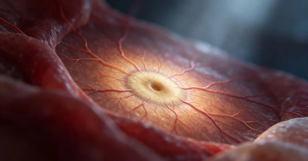
The macula sits in the middle of the retina, directly behind the lens. It is only a few millimetres across, yet it handles the fine, detailed part of your sight. At its centre lies a small pit called the fovea, where light-sensing cells called cones are packed most tightly.
Cones in the macula work best in good light and let you see colour and fine detail. The rest of the retina looks after side vision and helps you see in dim light. Our article on the difference between macula and retina explains how the two work together.
The macula is small, but it carries the vision you use for almost every detailed task.
What happens when the macula is damaged?
When the macula is damaged, the centre of what you look at can blur, darken or bend, while side vision usually stays. A person may still walk around a room easily but struggle to read a newspaper or see a grandchild’s face clearly.
Damage often builds slowly, so the better eye quietly makes up for the weaker one. Many people only notice a problem when they cover one eye by chance. Long-standing diabetic retinopathy can also reach the macula when fluid leaks from weakened blood vessels.
A change in central vision, even in one eye, is worth checking rather than putting down to age.

Common diseases of the macula
Several conditions can affect the macula, and their symptoms overlap. The right treatment depends on the cause, which is why an examination comes first.
Age-related macular degeneration
Age-related macular degeneration (AMD) slowly damages sharp central vision and is most common after the age of 60. Almost everyone with AMD starts with the dry form, in which small yellow deposits called drusen build up under the macula.
In the wet form, fragile new blood vessels grow under the macula and leak blood and fluid. It affects about 10 percent of people with AMD but causes most of the vision loss linked to the condition. Our retina team plans AMD treatment only after a full examination and scan.
Diabetic macular oedema
In people with diabetes, leaking blood vessels in the retina can make the macula swell with fluid. Central vision then looks blurred or washed out. Diabetic retinopathy treatment aims to reduce this swelling and protect the vision that remains, so tell both your eye doctor and your physician about any change.
Macular hole and macular pucker
A macular hole is a small break in the macula. A macular pucker is a thin layer of scar-like tissue that wrinkles its surface. Both can make straight lines look bent or leave a blurred patch in the centre, and where vision is affected a retina surgeon may discuss epiretinal membrane treatment.
Different macular diseases can feel alike, so the cause has to be confirmed before any treatment is planned.
Early signs of macular trouble
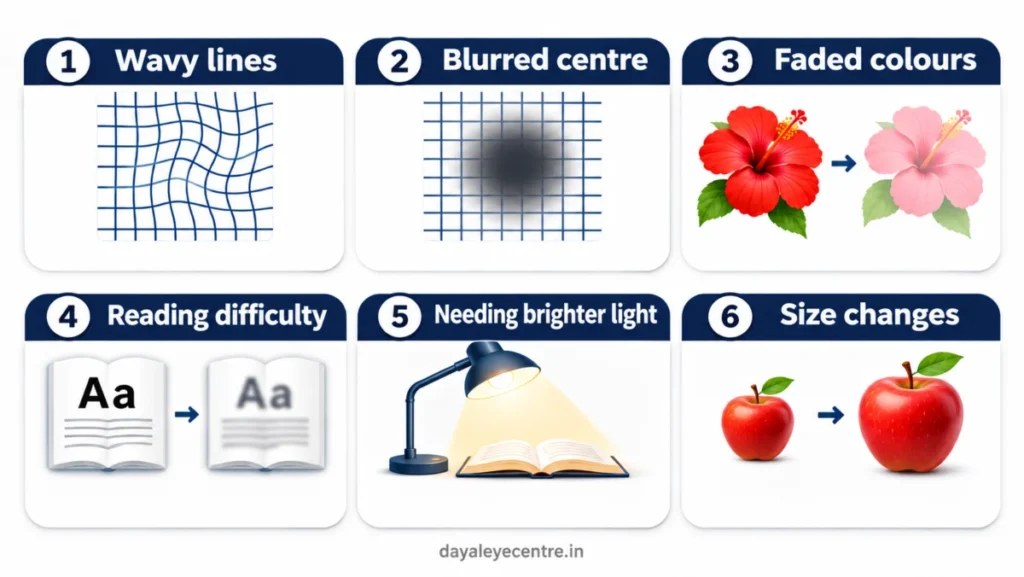
Macular problems often start quietly. Watch for:
- Straight lines that look wavy or bent
- A blurred or dark patch in the centre of vision
- Colours that look faded
- Difficulty reading or recognising faces
- Needing brighter light for close work
- Objects that look larger or smaller in one eye
Check one eye at a time, because the better eye can hide a change in the other. Small changes noticed early give your eye doctor more options.
How eye doctors check the macula
A macular check starts with a vision test and a dilated examination, where drops widen the pupil so the doctor can see the back of the eye. An OCT scan then builds cross-section images of the macula, layer by layer, without touching the eye. A digital retinal camera records photographs so changes can be compared at later visits.
At home, an Amsler grid, a printed square of straight lines with a dot in the middle, helps you spot early distortion. Information on age-related macular degeneration advises calling your eye doctor promptly if the lines look wavy.
Clinic scans and a simple home grid work best together.
When to see an eye doctor
Book an examination if you notice any early sign, if you have diabetes or if AMD runs in your family. A grandparent who has started holding the newspaper at odd angles to find a clear spot should be checked soon.
Seek in-person eye care straight away for sudden loss or dimming of vision, a curtain or shadow across vision, or a sudden shower of new floaters or flashes. Call Dayal Eye Centre on +91 81786 39701 during opening hours, or go to the nearest hospital emergency department.
Retinal problems are time-sensitive, so a sudden change deserves a same-day visit.
This article is general information. An ophthalmologist needs to examine your eyes to advise on your own case.
Protecting your central vision
The macula gives you sharp central vision, and most of its diseases show early signs such as wavy lines or a blurred centre. Checking one eye at a time and keeping up regular examinations makes early changes easier to catch. If anything has changed, book a retina check-up at Dayal Eye Centre in Sector 15, Gurugram.
Frequently asked questions
Can the macula heal on its own?
Some swelling settles once its cause is treated. Damage from AMD cannot be reversed, so treatment aims to slow it and protect the vision you have.
Is macular degeneration the same as blindness?
No. AMD affects central vision, and side vision usually remains, so most people can still move around independently.
Can young people have macular disease?
Yes. Diabetes, high short-sightedness and inherited conditions can affect the macula earlier, although AMD itself is uncommon before 55.
Does diet help the macula?
Green leafy vegetables contain lutein and zeaxanthin, natural pigments found in the macula. A balanced diet supports eye health but does not replace check-ups.
How often should the macula be checked?
Your eye doctor will advise based on your age, diabetes and family history. Adults over 60 are commonly advised a dilated eye examination about once a year.
Does an OCT scan touch the eye?
No. You rest your chin on a support and look at a light for a few seconds while the machine takes its images.
Does smoking affect the macula?
Smoking is a known risk factor for AMD. Stopping is one of the most useful steps you can take for your eyes.