Vitrectomy is the surgery we recommend when a problem deep inside the eye — bleeding, scar tissue, a macular hole or a detached retina — is threatening your sight and cannot be corrected with drops, injections or laser alone. At Dayal Eye Centre, our fellowship-trained retina surgeons use small-gauge, stitch-free vitrectomy (also called pars plana vitrectomy, or PPV) to gently remove the eye’s vitreous gel — the clear jelly that fills the back of the eye — relieve the pull on your retina, and repair the damage from within.
Accredited eye hospital — audited safety and infection-control protocols
1.2 lakh+
Surgeries performed across the centre
23+ yrs
Surgical experience in Gurgaon
Fellowship
Trained retina specialists leading every vitrectomy
Dedicated
Modular retina theatre with an on-site in-patient ward
Not sure if vitrectomy is what your eye needs?Consult with our doctors for a detailed retinal evaluation.
Candidacy
Is Vitrectomy the Right Surgery for You?
Vitrectomy is not a first step — it is the treatment we turn to when the vitreous gel is clouded by blood, is pulling on the retina, or is blocking access to a problem that needs repair. Your surgeon recommends it only after a detailed retinal examination and scans confirm it is the safest way to protect or restore your vision. We commonly perform vitrectomy for:
Retinal detachment
Especially complex, recurrent or tractional detachments, where the retina has pulled away from the back of the eye (see our retinal detachment care).
Advanced diabetic eye disease
Diabetic retinopathy with vitreous haemorrhage (bleeding into the gel) or scar tissue dragging on the retina.
Macular hole and epiretinal membrane
Also called macular pucker — fine membranes are peeled to help the central retina return to shape (macular hole & ERM).
Floaters that do not settle over time and genuinely interfere with daily life.
Other retinal emergencies
Eye infection (endophthalmitis), serious eye injury, a dislocated natural lens or intraocular lens, or lens fragments left behind after cataract surgery.
Age alone rarely rules you out — many of our vitrectomy patients are older adults, and people living with diabetes can be excellent candidates once their blood sugar, blood pressure and eye pressure are assessed. What matters is the condition of your retina, your general health and how well your eye is likely to heal, all of which we establish before advising surgery.
And if a gentler option — an injection, laser, or careful observation — can achieve the same result, we will tell you so honestly.
The Procedure
What Happens During Vitrectomy Surgery at Dayal Eye Centre?
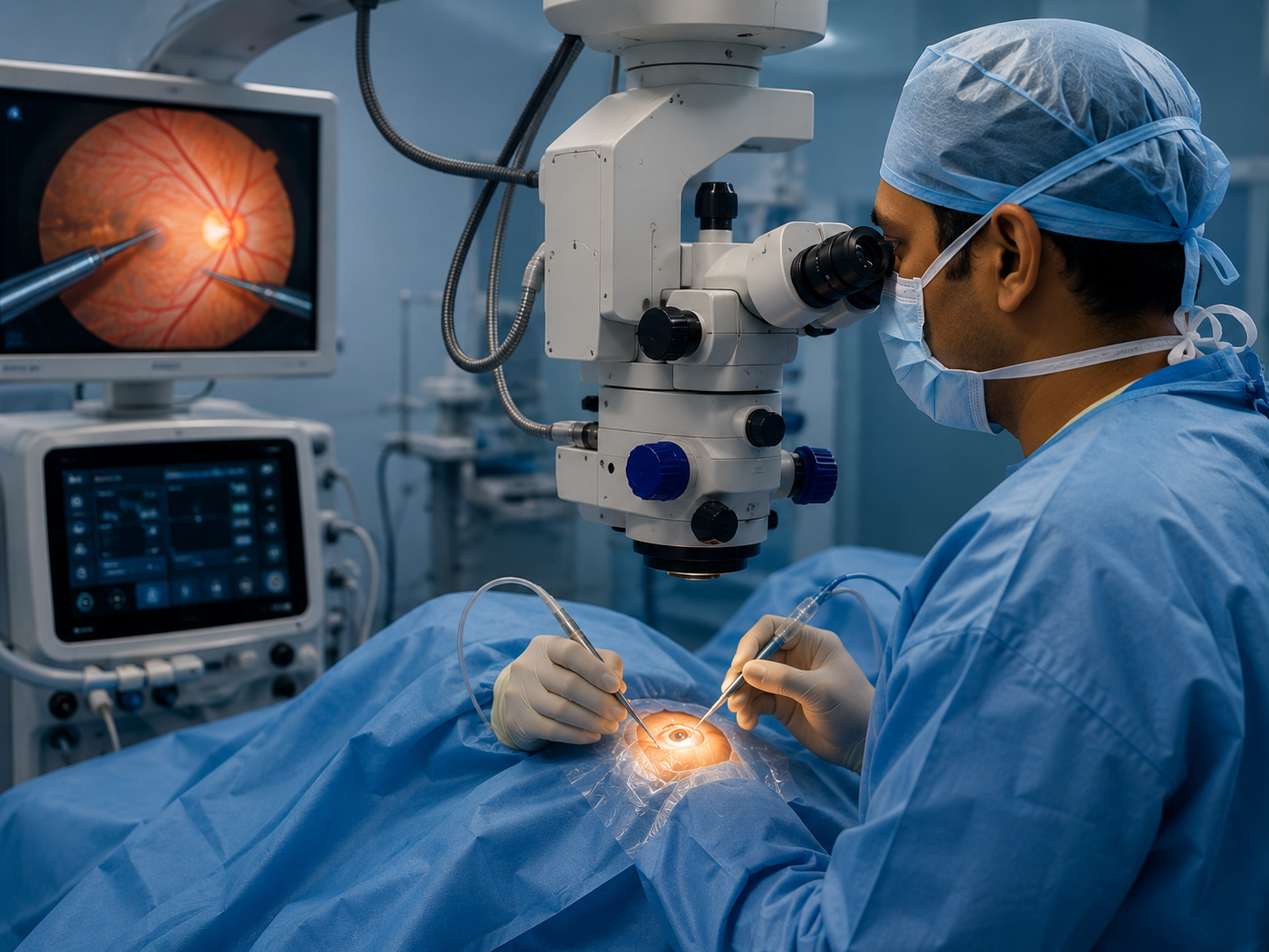
Modern vitrectomy is a microincision, stitch-free procedure. Through three openings smaller than a millimetre in the white of the eye (the sclera), your surgeon inserts microscope-guided instruments — a fine light source, an infusion line that keeps the eye gently pressurised and stable, and a high-speed cutter that removes the vitreous gel. Once the gel is cleared, the surgeon can reach the retina directly to clear blood, remove scar tissue, peel delicate membranes, seal tears with laser or freezing treatment (cryotherapy), and flatten a detached retina.
To hold the retina in place while it heals, the surgeon may fill the eye with a temporary tamponade — a gas bubble, air, or silicone oil — chosen to suit your specific repair. A gas or air bubble is gradually absorbed and replaced by your eye’s own natural fluid; silicone oil is removed later in a minor follow-up procedure. Most vitrectomies at Dayal Eye Centre are performed as day-care surgery under local anaesthesia and take roughly 45 minutes to a couple of hours, depending on how complex the repair is.
1
Three sub-millimetre ports
Openings smaller than a millimetre are made in the white of the eye (the sclera) — no stitches needed in most cases.
2
Microscope-guided instruments
A fine light source, an infusion line that keeps the eye gently pressurised and stable, and a high-speed cutter that removes the vitreous gel.
3
Repair from within
With the gel cleared, your surgeon clears blood, removes scar tissue, peels delicate membranes, seals tears with laser or cryotherapy, and flattens a detached retina.
4
Tamponade to hold the retina
A gas bubble, air or silicone oil supports the retina while it heals. Gas and air absorb on their own; silicone oil is removed later in a minor follow-up procedure.
The Vitrectomy Techniques We Use
Your surgeon selects the approach that matches your diagnosis — and explains it to you before the day of surgery.
Vitrectomy approach
What it involves
Commonly used for
Small-gauge (23G / 25G / 27G) MIVS
Sutureless micro-incision vitrectomy through sub-millimetre ports
Most retinal repairs — supports a faster, more comfortable recovery
Vitrectomy with membrane peeling (ILM / ERM)
Removing wrinkled membranes from the surface of the macula
Clearing blood from the vitreous cavity to restore a clear view
Diabetic bleeds, retinal vein occlusion, trauma
Technology
What Technology and Diagnostics Support Your Surgery?
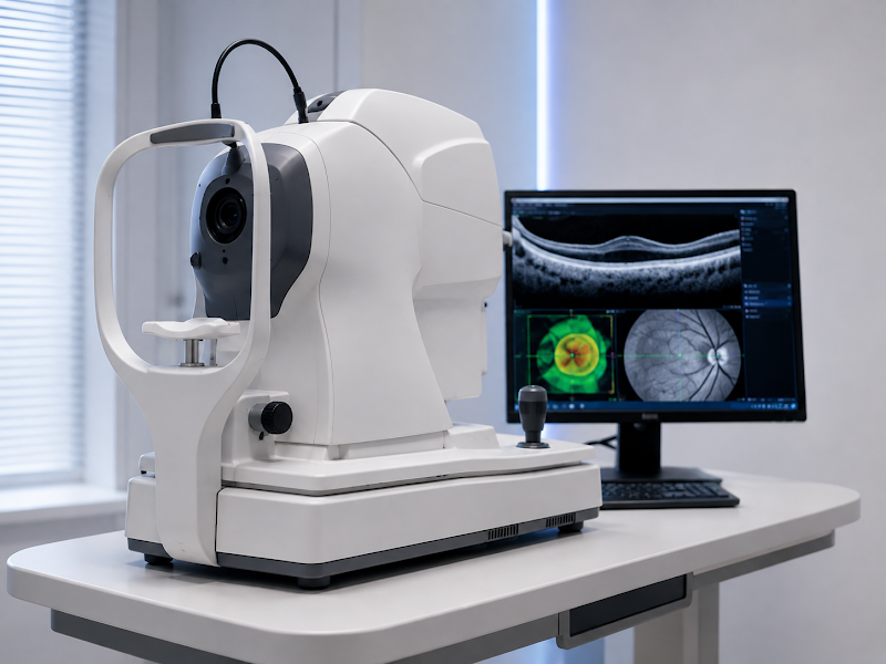
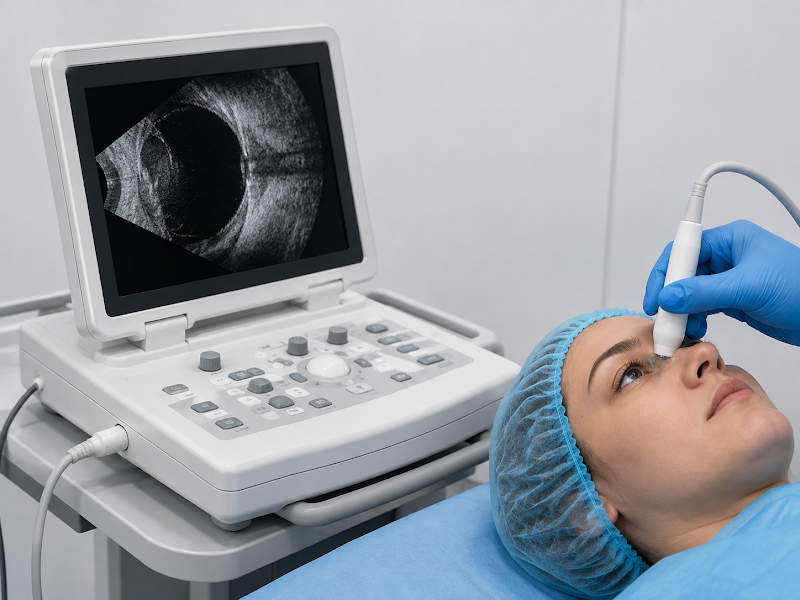
A vitrectomy is only as good as the planning behind it. Before surgery, we map your retina in detail using Optical Coherence Tomography (OCT) for cross-sectional images of the macula, B-scan ultrasound when a dense bleed blocks the view inside the eye, and fluorescein angiography to study the blood flow in your retina.
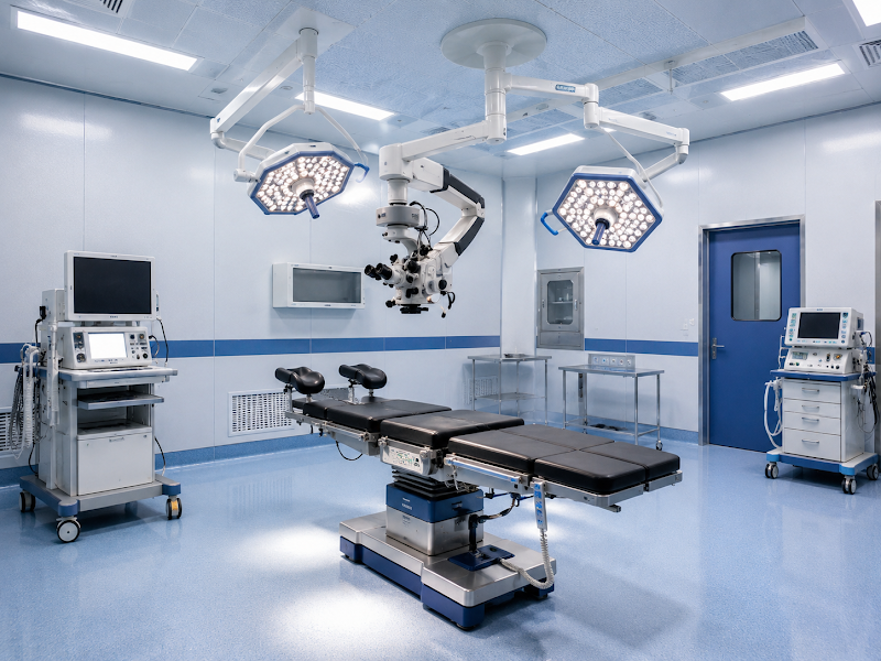
Surgery is carried out in one of our three modular operation theatres — including a theatre dedicated to retina work — with an on-site in-patient ward for cases that need overnight observation. Having advanced diagnostics and surgery under one roof means fewer referrals and a faster path from diagnosis to treatment. Not sure where to start? A detailed eye check-up is the first step.
Optical Coherence Tomography (OCT)
Cross-sectional images of the macula that show holes, traction, swelling and thinning layer by layer — the scan that shapes your surgical plan.
B-scan ultrasound
Used when a dense vitreous bleed blocks the view inside the eye, so we can still assess whether the retina is attached before surgery.
Modular retina theatre
One of three modular operation theatres, including a theatre dedicated to retina work, with an on-site in-patient ward for overnight observation.
Recovery from vitrectomy is a gradual, step-by-step process, and being prepared for it makes it far less daunting. In the first few days your vision is usually blurry and the eye may feel gritty or mildly sore; this settles as the eye heals. If a gas bubble was placed, you may see a dark, wobbling line or bubble in your vision that slowly shrinks and disappears as the gas is absorbed — typically over about two to eight weeks, depending on the type of gas used.
Head positioning
If a bubble was used, your surgeon may ask you to keep your head in a specific position — often face-down — for several days to a few weeks, so the bubble presses gently on the healing area of the retina. It is the hardest part of recovery for many patients, which is exactly why our team helps you plan for it before surgery, with practical tips and positioning support.
Vision timeline
Many people notice clearer vision within a few weeks, but full visual recovery can take a few months, and longer in complex cases. Some patients experience temporary double vision or a sense of spatial disorientation in the early weeks as the eye adjusts.
If you still have your natural lens, it is common for a cataract to develop in the operated eye within a year or so — this is an expected part of healing and is easily treated later with routine cataract surgery.
An important safety rule
While a gas bubble is in the eye you must not fly, travel to high altitude, or have certain anaesthetics — the gas can expand and dangerously raise the pressure inside your eye. Your surgeon will confirm exactly when it is safe again.
Most patients return to light daily activities within a few days, while avoiding heavy lifting, straining, rubbing the eye and swimming until cleared. We schedule regular follow-ups to track your healing closely and to catch any problem early.
Outcomes
What Are the Success Rates and Risks of Vitrectomy?
Vitrectomy is one of the most refined procedures in modern eye surgery, and for many conditions its results are very good. Published studies report single-surgery macular hole closure rates of roughly 90–93%, rising further with today’s membrane-peeling techniques, and about three-quarters of patients gain two or more lines of vision after a successful macular hole repair. For retinal detachment, pars plana vitrectomy reattaches the retina in roughly 9 out of 10 eyes with a single operation, and in close to all eyes after further surgery where it is needed. Because these figures come from the wider research literature, they describe what is typically achievable across many patients — not a promise about your individual eye.
90–93%
Single-surgery macular hole closure reported in published studies
~75%
Of patients gain two or more lines of vision after successful macular hole repair
9 in 10
Eyes reattached with a single pars plana vitrectomy for retinal detachment
1–3%
Reported rate of new or recurrent detachment in these studies
Like any surgery, vitrectomy carries risks
The main ones include cataract formation (common, and treatable), raised eye pressure, infection, bleeding, and a small chance of a new or recurrent retinal detachment — reported in roughly 1–3% of cases in these studies. Your final result depends on the condition being treated, how long it was present before surgery, the health of your macula and optic nerve, and how well your eye heals.
Individual results vary based on your eye condition, its severity and duration, your age, overall eye health and healing response. Our surgeons explain the specific benefits and risks for your eye before you decide on surgery.
Sources: macular hole closure — Mass Eye & Ear retina outcomes, PMC5932482, Retina Today 2024; vision gain — Kelly & Wendel via PMC5932482; reattachment rates — Mass Eye & Ear (PPV 94.6% single surgery) and published series 82–95%; new or recurrent detachment — Ophthalmology Advisor, Nature 2017.
Cost & Payment
How Much Does Vitrectomy Surgery Cost in Gurgaon?
There is no single fixed price for vitrectomy, because the cost depends on the condition being treated and how complex the repair is. As a guide, vitreo-retinal surgery at Dayal Eye Centre typically starts from around ₹51,000 per eye, with your exact estimate confirmed only after an examination — this is a starting-from figure, not a fixed quote. We share a clear, itemised estimate before surgery, so there are no surprises. The main factors that shape your cost are:
Factor affecting cost
Why it matters
Condition & complexity
A straightforward vitreous bleed usually costs less than a complex or recurrent detachment repair
Tamponade used
Silicone oil needs a second, minor procedure later to remove it
Type of anaesthesia
Local (day-care) versus general anaesthesia
One or both eyes
Bilateral surgery is planned and priced separately
Combined procedures
Vitrectomy is sometimes combined with cataract surgery in the same sitting
From ₹51,000per eye · starting-from figure, not a fixed quote
Vitrectomy for a medical retinal condition is usually covered by health insurance. We offer cashless treatment across major insurer and TPA networks, Easy EMI via Savein, and support government and PSU schemes including CGHS, ECHS, Ayushman Bharat and Haryana Government schemes. Our counsellors help you plan the finances before surgery.
Want a clear estimate for your eye?Consult with our doctors and we’ll confirm your options after an examination.
Why Dayal Eye Centre
Why Choose Dayal Eye Centre for Vitrectomy in Gurgaon?
Retina surgery rewards experience, and at Dayal Eye Centre your vitrectomy is performed by fellowship-trained retina specialists within a fully equipped, NABH-accredited eye hospital — not a general set-up. With diagnostics, three modular operation theatres and an in-patient ward under one roof — plus cashless insurance and Easy EMI options — we make complex retina care both accessible and reassuring. Whether your surgery is planned or urgent, you are looked after by senior hands from the first scan to the final follow-up.
Leads our retina service as a vitreoretinal surgeon. She completed fellowships in vitreo-retina surgery at H.V. Desai Eye Hospital, Pune and medical retina at Shroff Eye Centre, Delhi, with 8+ years of surgical experience.
Our Director is one of Gurgaon’s most experienced eye surgeons, with 23+ years of practice and more than 80,000 surgeries to his name — supported by a full team of sub-specialists.
Meet the full team — senior sub-specialists across retina, cataract, cornea, glaucoma and oculoplasty.
Book Your Vitrectomy Consultation at Dayal Eye Centre
Concerned about your retina, or advised to have vitrectomy elsewhere and want a second opinion? Consult with our doctors — senior retina specialists, under one roof.
808 B, Galaxy Road, Sector 15 Part 2, Near 32nd Avenue, Gurgaon, Haryana 122001
Phone / WhatsApp
+91 81786 39701
Email
wecare@dayaleyecentre.in
Hours
Monday–Saturday, 8:30 AM – 7:30 PM · Sunday closed
Getting here
Beside 32nd Avenue, off NH-48 · ~25 min from Delhi · nearest metro IFFCO Chowk (Yellow Line)
FAQs
Frequently Asked Questions About Vitrectomy Surgery
The questions patients ask us most before deciding on vitrectomy surgery in Gurgaon.
Vitrectomy is carried out under anaesthesia, so you should not feel pain during the surgery itself. Afterwards, most patients feel only mild discomfort, grittiness or a watery sensation for a few days, which is managed with prescribed drops and simple pain relief. Tell your surgeon if you have any severe or worsening pain, as that is uncommon and should be checked.
Vision is usually blurry at first and clears gradually. Many people notice improvement within a few weeks, but full visual recovery can take a few months — and longer for complex conditions. If a gas bubble was used, your vision stays blurry until the bubble absorbs over about two to eight weeks.
Only if a gas or air bubble was placed in your eye. In that case your surgeon may ask you to keep your head face-down (or on a particular side) for several days to a few weeks, so the bubble supports the healing retina. Not every vitrectomy needs positioning — your surgeon will tell you what applies to your case, and our team helps you prepare for it.
Not while a gas bubble is still in your eye. Air travel and high altitude can cause the gas to expand and dangerously raise the pressure inside the eye. You can usually fly once your surgeon confirms the bubble has fully absorbed. If silicone oil or no bubble was used, flying restrictions are different — always check with your surgeon first.
If you still have your natural lens, it is common to develop a cataract in the operated eye within a year or so of vitrectomy. This is an expected part of healing rather than a complication, and it is easily treated later with routine cataract surgery.
Yes — in fact, advanced diabetic eye disease is one of the most common reasons for vitrectomy. Good control of your blood sugar and blood pressure, along with an assessment of your eye pressure and general health, helps your eye heal well. Our retina team has a special focus on diabetic retinopathy.
Success rates are high for many conditions — for example, published studies report macular hole closure of around 90–93% and retinal reattachment in roughly 9 out of 10 eyes with a single operation. Your own outcome depends on the condition treated, how early it is addressed and how your eye heals, so results vary from person to person.
Age alone rarely rules you out. Many vitrectomy patients are older adults, and what matters most is the condition of your retina and your overall health. A detailed evaluation tells us whether surgery is the safest way to protect your vision.
Vitrectomy for a medical retinal condition is usually covered by health insurance. We offer cashless treatment across major insurer and TPA networks, plus Easy EMI and support for CGHS, ECHS, Ayushman Bharat and Haryana Government schemes.
Medically reviewed by Dr. Neelam Khatwani, MBBS, MS, DNB, FVRS — Vitreoretinal Surgeon, Dayal Eye Centre · Last reviewed: 31 July 2026 · Editorial Policy

 Vision Correction
 Cataract
 Retina
 Glaucoma & Cornea
 Paediatric & Speciality
 Oculoplasty