NABH Accredited 20+ Years 1.2 Lakh+ Surgeries International Patients Mon–Sat · 8:30 AM–7:30 PM +91 81786 39701
NABH Accredited
Dayal Eye Centre
Contact
Book an AppointmentHow would you like to go ahead?
Self BookingPick your doctor, date and slot. Booking RequestLeave your number — our counsellors call you. Mon–Sat 8:30 am – 7:30 pm · Sunday closed
NABH · Gurugram

Need a second opinion on your eyes?

Leave your number. A specialist calls you back today.

Available now

Call us from your phone

Copy the number, or leave your details and we will call you back.

+91 81786 39701

Mon–Sat · 8:30 AM–7:30 PM · Sector 15, Gurugram
Understanding Your Eyes

Eye Care Glossary

Plain-English explanations of the eye conditions, tests and treatments you'll hear about at Dayal Eye Centre — the medical term, and what it actually means for you.

Jump to
A B C D E F G H I J K L M N O P Q R S T U V W X Y Z
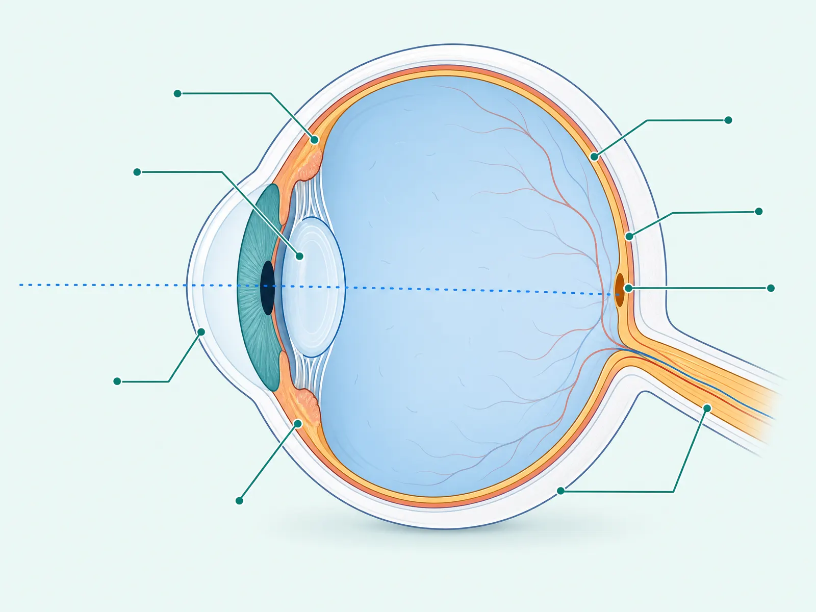
Cross-section diagram of the human eye with major parts indicated
How the Eye Works

A quick map of your eye

Light enters through the cornea, passes the pupil and lens, and focuses on the retina — whose central macula gives you sharp detail. The optic nerve carries it all to the brain.

Illustration — simplified for education.

53 terms — conditions, anatomy, tests, procedures & optics
#

6/6 Vision

Test

The benchmark for "normal" distance sight: you read at 6 metres what a normal eye reads at 6 metres. It describes clarity at distance only — not your overall eye health.

A
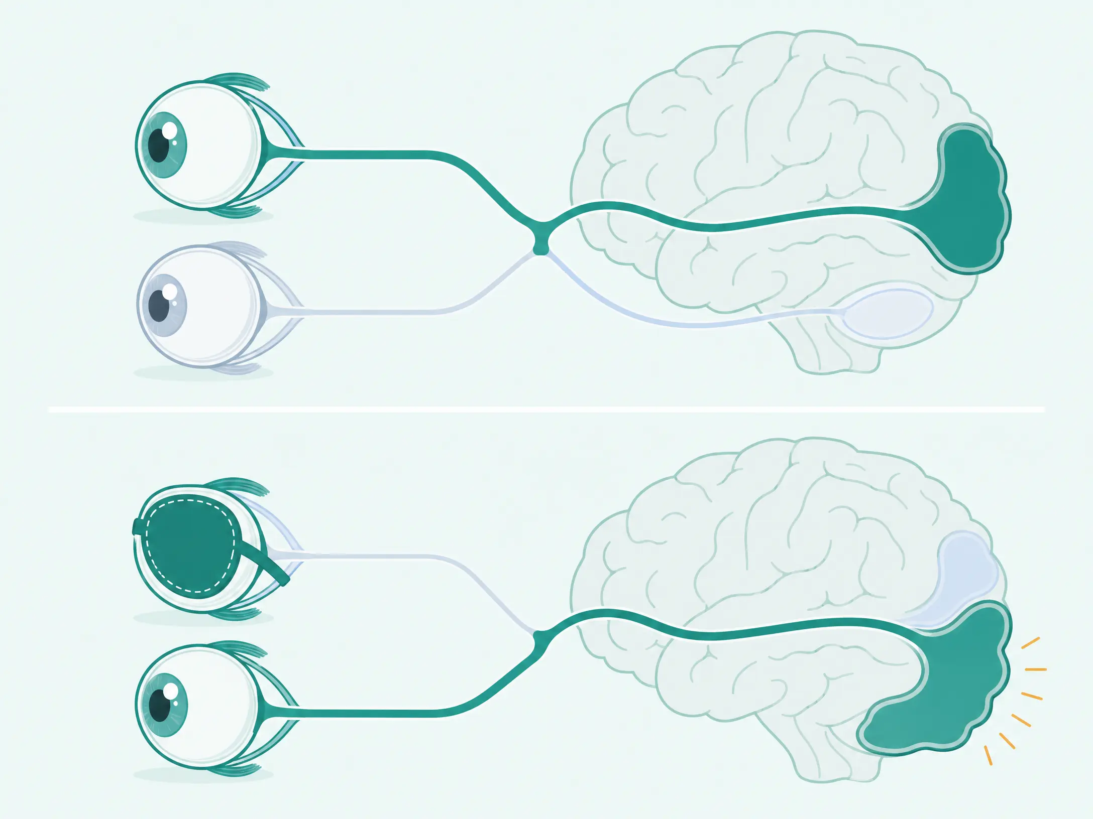
Diagram of amblyopia and patching therapy strengthening the weaker eye

Amblyopia Lazy eye

Condition

When one eye and the brain don’t develop a strong connection in childhood, so that eye sees weaker even with glasses. Patching the stronger eye trains the weaker one — it works best when treated early.

See also Squint
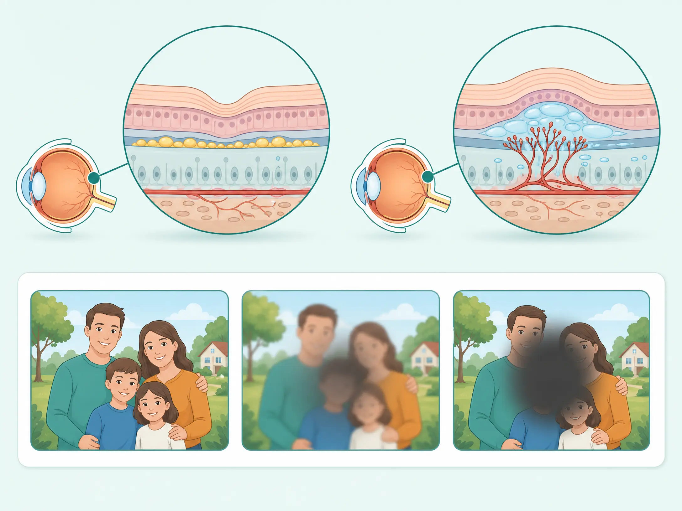
Diagram of dry and wet age-related macular degeneration

AMD Age-related macular degeneration

Condition

Ageing damage to the macula, the central part of the retina. "Dry" AMD is slow and driven by deposits called drusen; "wet" AMD is faster, from leaky new vessels. It affects central vision while side vision is preserved.

See also Macula Drusen Retina

Astigmatism

Condition

A cornea (or lens) shaped more like a rugby ball than a football, so light focuses at more than one point. The result is blur at all distances, often with some eye strain.

Axis

Optics

The number from 0 to 180 on your glasses prescription that tells the direction of your astigmatism correction. It only matters alongside a cylindrical (CYL) value.

C
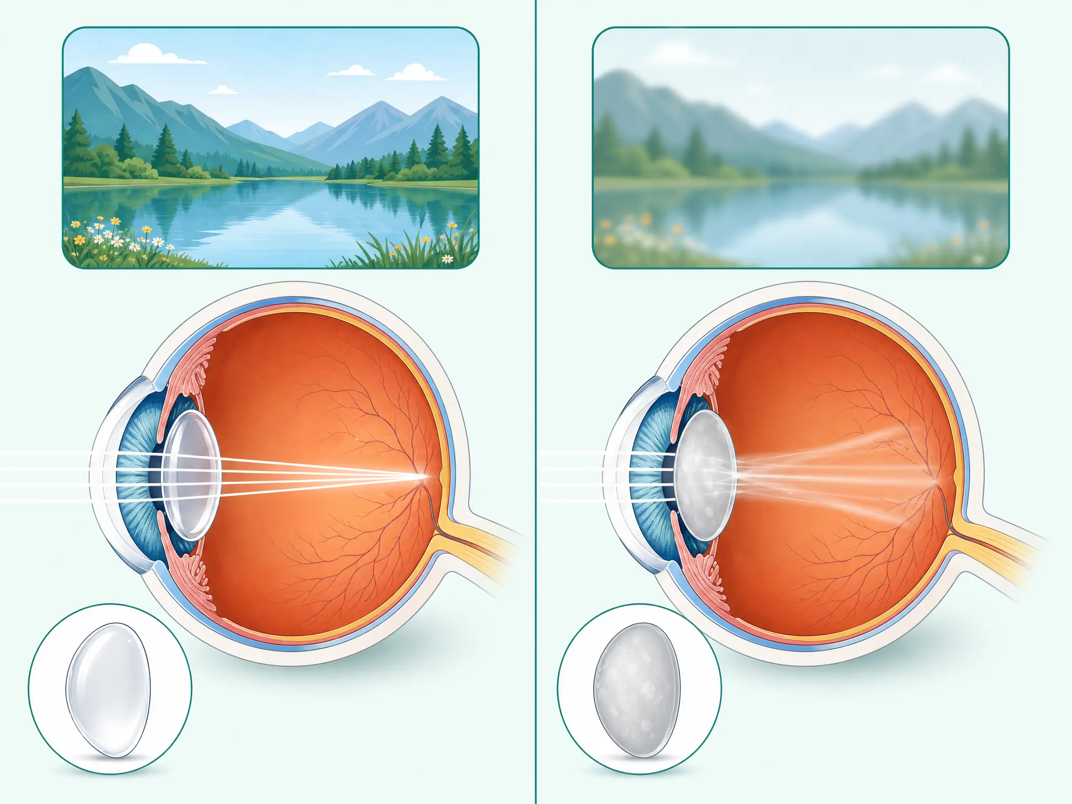
Comparison of an eye with a clear lens and an eye with a cataract

Cataract Motiyabind

Condition

Clouding of the eye’s natural lens, usually with age, making vision dim, hazy or glary. It is corrected by replacing the cloudy lens with a clear artificial one (an IOL) during surgery.

Ciliary Body

Anatomy

The ring of muscle behind the iris that changes the shape of the natural lens to focus, and produces the fluid that fills the front of the eye.

See also Lens Glaucoma

Cornea

Anatomy

The clear front dome of the eye. It does most of the eye’s focusing, which is why reshaping it with a laser (LASIK, SMILE, PRK) can correct your vision.

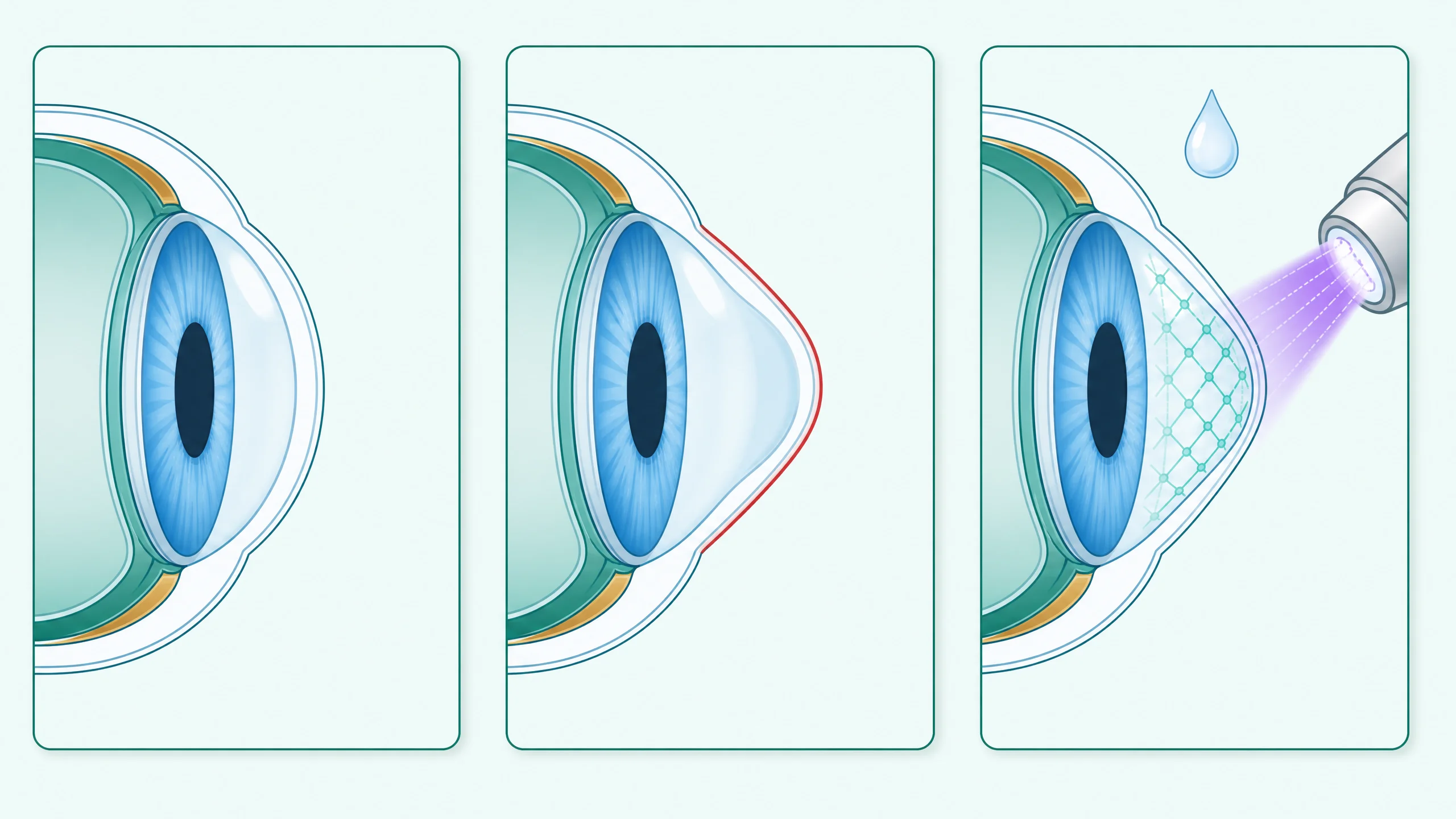
Corneal Cross-Linking C3R

Procedure

A treatment that strengthens a weak, thinning cornea using vitamin-B2 drops and controlled ultraviolet light. It halts keratoconus from getting worse — it stabilises, rather than reverses.

Cylindrical Power CYL

Optics

The part of your prescription that measures the strength of astigmatism. It works together with the AXIS value, which sets its direction.

D
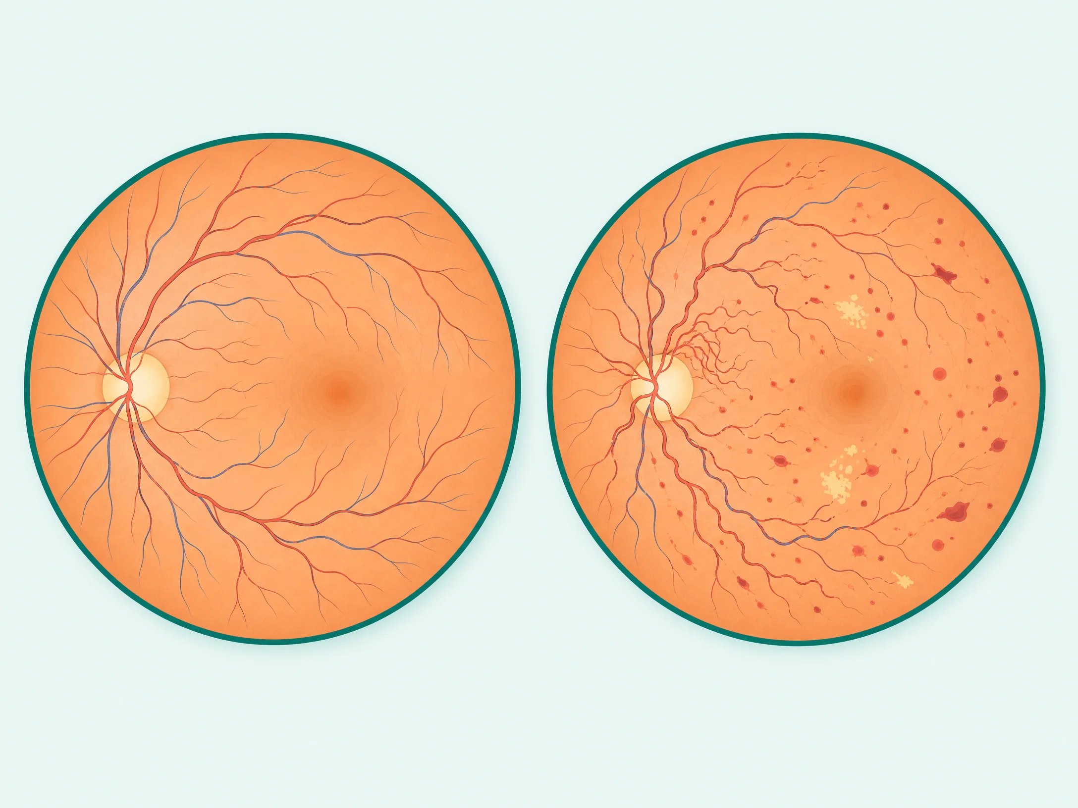
Comparison of a healthy retina and a retina with diabetic retinopathy

Diabetic Retinopathy

Condition

Damage to the retina’s tiny blood vessels caused by diabetes. It often has no early symptoms, so every person with diabetes needs a yearly dilated retina check.

See also Retina OCT

Drusen

Condition

Small yellow deposits that build up under the retina with age. A few are common; many can be an early sign of dry AMD.

See also AMD Macula

Dry Eye

Condition

When the tear film is unstable or your eyes don’t make enough good-quality tears, causing burning, grittiness and moments of blur. Very common with screens and air-conditioning.

See also Tear Film
E

EDOF Extended depth-of-focus IOL

Optics

A type of artificial lens that stretches one continuous zone of focus from distance through to arm’s length — good for distance and screen use with fewer visual side-effects than trifocals.

F

FLACS Femtosecond laser-assisted cataract surgery

Procedure

Cataract surgery in which key steps are performed by a computer-guided laser rather than by hand, for added precision on selected eyes.

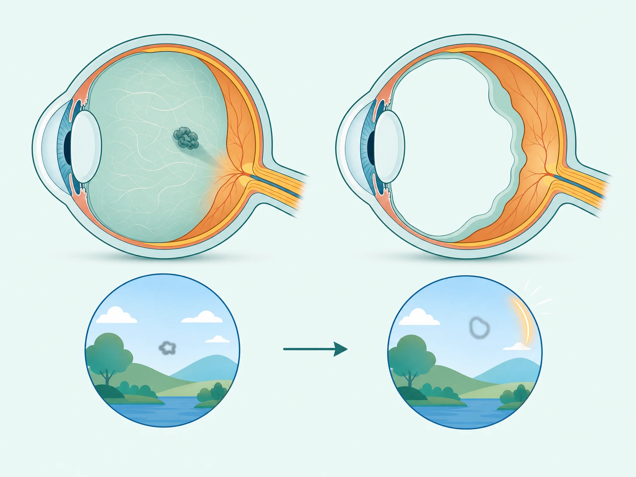
Diagram showing why floaters appear and what they look like

Floaters

Condition

Specks, threads or cobwebs that drift across your vision — shadows cast by clumps in the vitreous gel. Usually harmless, but a sudden shower of floaters or flashes needs a same-day check.

G
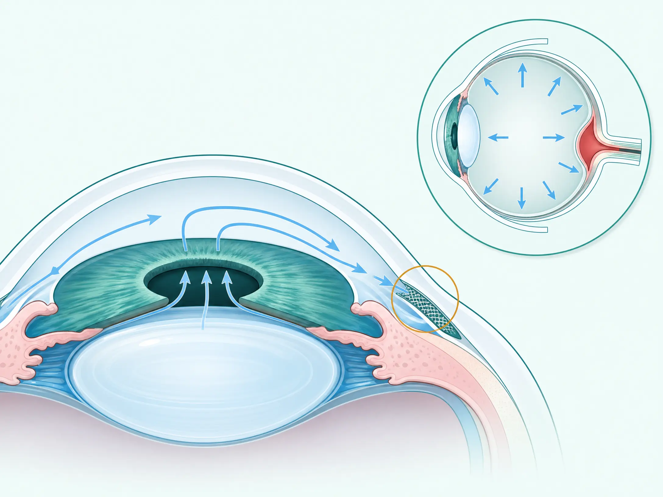
Diagram of eye fluid drainage and pressure affecting the optic nerve in glaucoma

Glaucoma

Condition

A group of conditions where raised pressure inside the eye damages the optic nerve, silently narrowing vision from the sides inward. Early detection protects the sight you have.

H

Hyperopia Farsightedness

Condition

A shorter-than-average eye, so light focuses behind the retina. Near objects blur first, and effort is needed to keep things sharp.

I
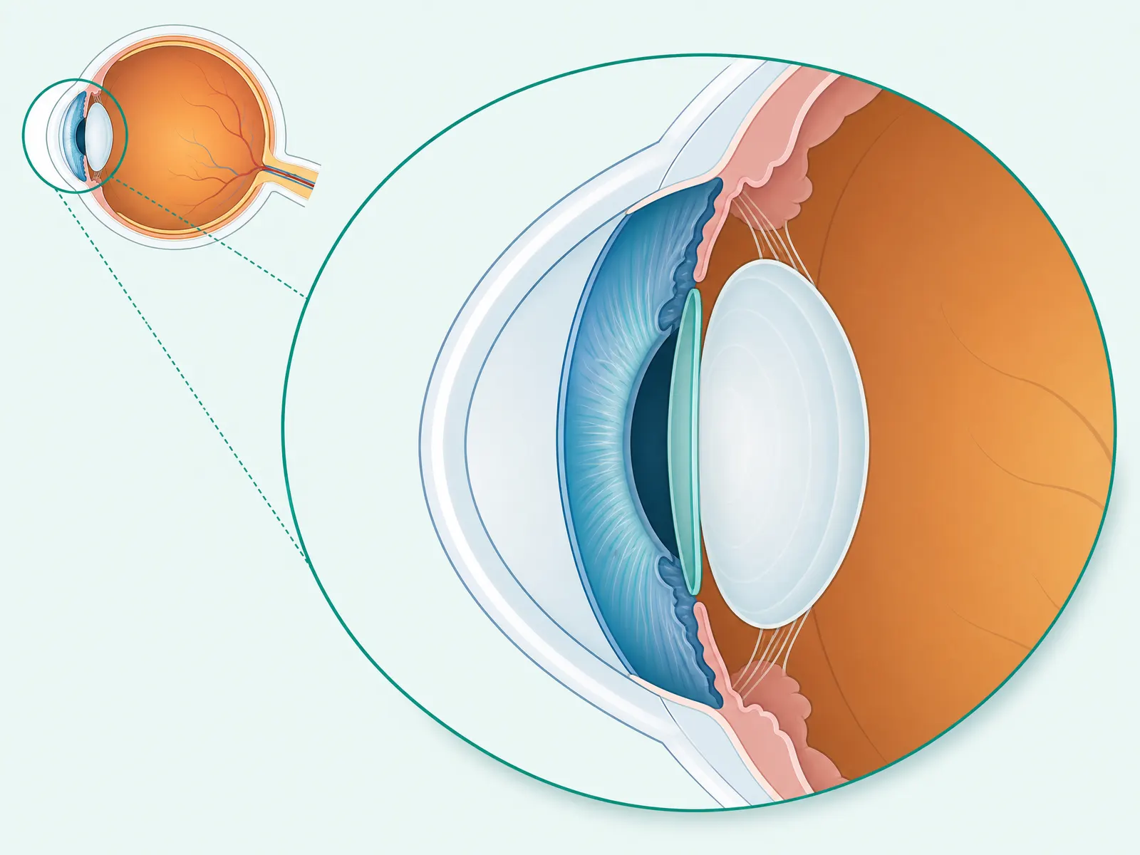
Diagram showing an implantable collamer lens positioned behind the iris

ICL Implantable Collamer Lens

Procedure

A thin corrective lens placed inside the eye, behind the iris, while your natural lens stays in place. A good option for high powers or corneas too thin for LASIK — and it’s removable.

See also Phakic IOL Iris
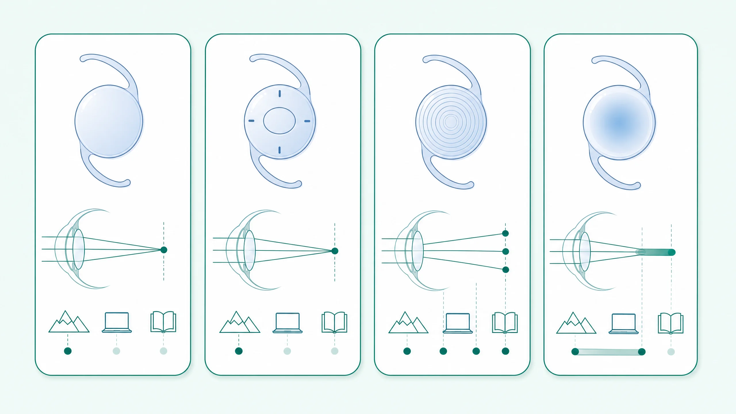
Diagram of four intraocular lens types and their focus ranges

IOL Intraocular lens

Optics

The clear artificial lens that replaces your cloudy natural lens during cataract surgery. Choices include monofocal, toric, multifocal/trifocal and EDOF designs.

Iris

Anatomy

The coloured ring of the eye. Its muscles open and close the pupil to control how much light gets in.

See also Pupil
K
Diagram of a normal cornea, keratoconus, and corneal cross-linking treatment

Keratoconus

Condition

A cornea that gradually thins and bulges into a cone, distorting vision. Caught early it can be stabilised with corneal cross-linking.

L
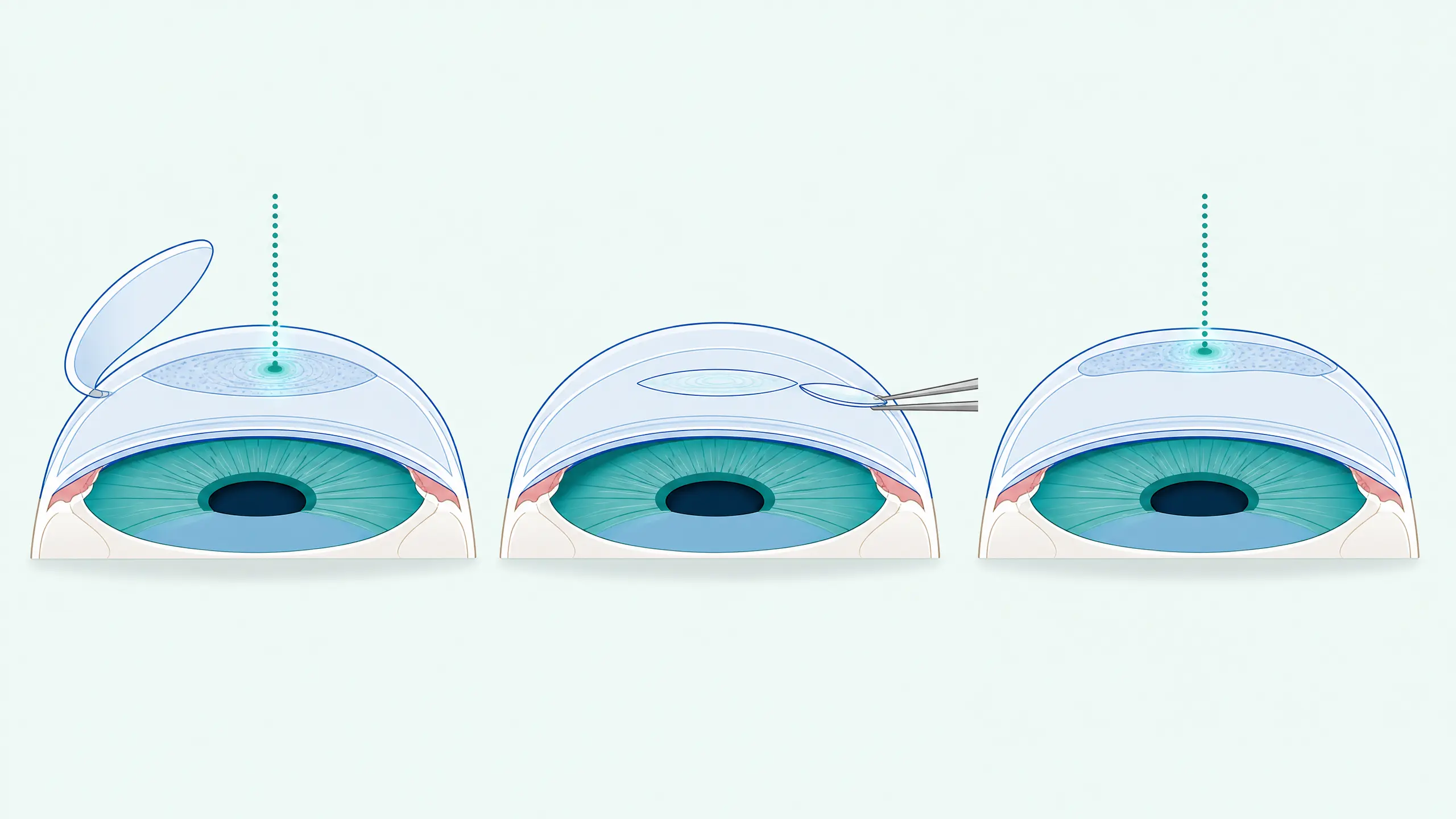
Diagram comparing LASIK, SMILE and PRK corneal procedures

LASIK

Procedure

Laser vision correction in which a thin hinged flap is created in the cornea and a laser reshapes the layer beneath — a quick recovery for suitable eyes.

See also Cornea PRK SMILE

Lens Crystalline lens

Anatomy

The natural lens sitting behind the pupil that fine-tunes focus. It clouds into a cataract with age and stiffens with age (presbyopia).

M

Macula

Anatomy

The small central part of the retina responsible for sharp, detailed, straight-ahead vision — reading, faces, fine work.

See also Retina AMD

Monofocal

Optics

An artificial lens set for a single focus, usually distance. Clear far vision, with reading glasses for near — the most widely used IOL.

Multifocal / Trifocal

Optics

An artificial lens with rings that provide several focal points — far, screen and near — reducing dependence on glasses after cataract surgery.

See also IOL EDOF Monofocal

Myopia Nearsightedness

Condition

A longer-than-average eye, so light focuses in front of the retina and distant objects blur. Common, rising in children, and correctable with glasses, lenses or laser.

O
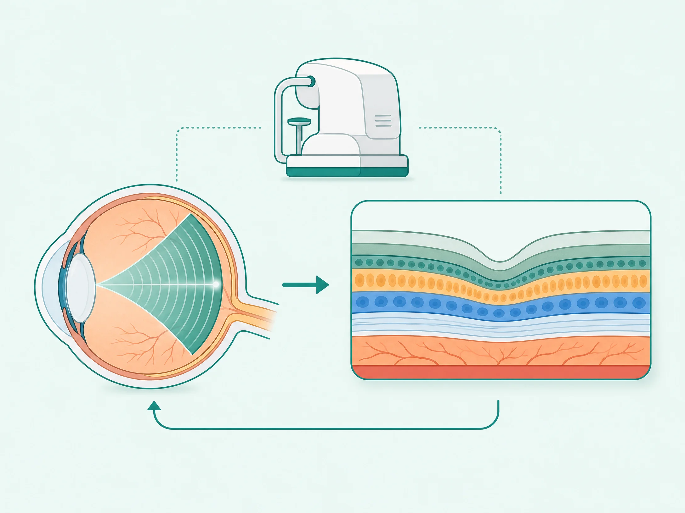
Diagram explaining how an OCT scan images the retinal layers

OCT Optical coherence tomography

Test

A painless light scan that builds a cross-section of the retina, layer by layer — no touch involved. It detects macula and optic-nerve changes very early.

Optic Nerve

Anatomy

The cable that carries visual signals from the retina to the brain. Damage to it — as in glaucoma — cannot be reversed, so protecting it matters.

See also Glaucoma Retina
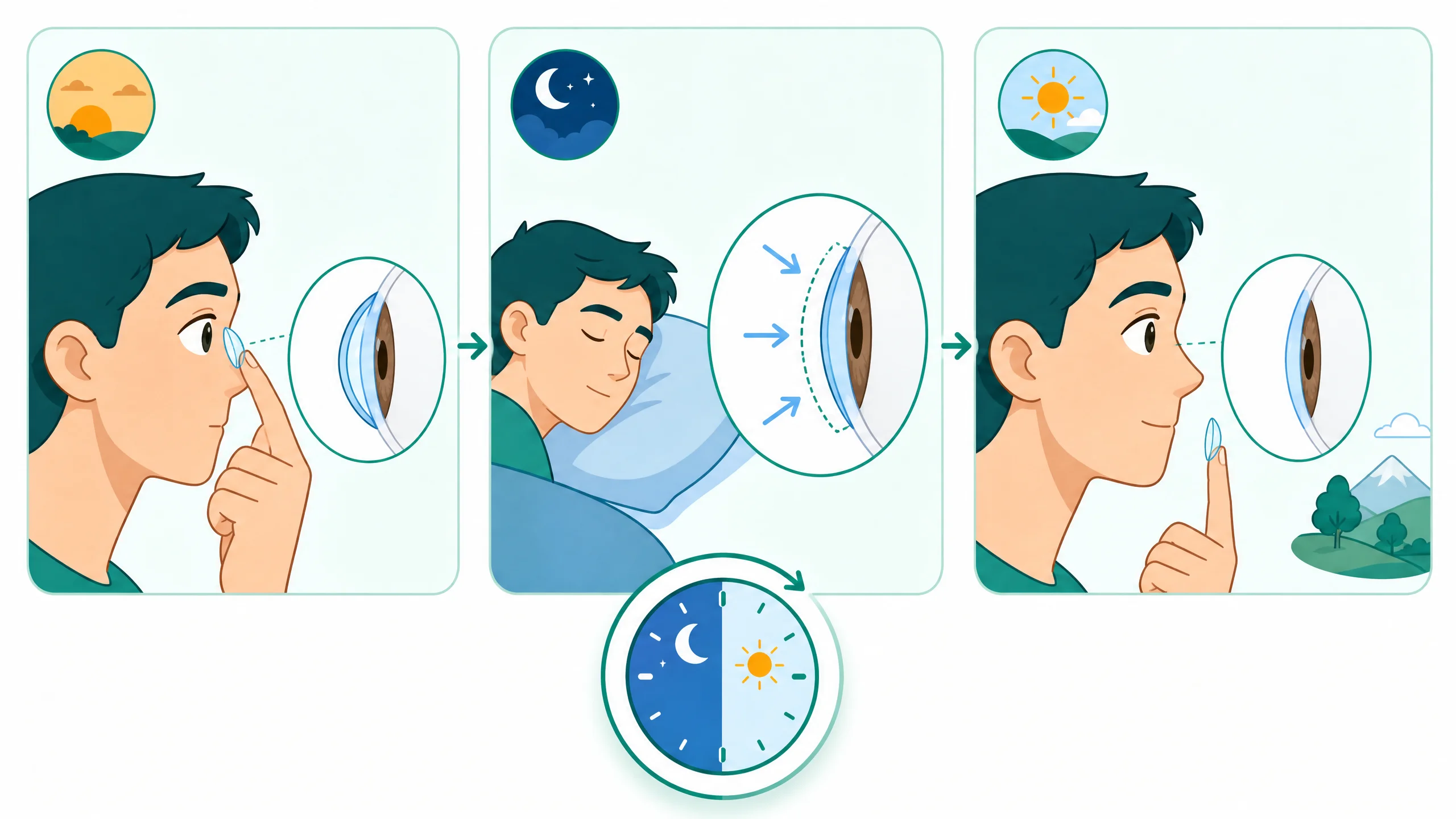
Diagram of the overnight orthokeratology vision correction cycle

Ortho-K Orthokeratology

Procedure

Special lenses worn overnight that gently reshape the cornea, giving lens-free clear vision through the day. The effect is temporary and reversible — nightly wear maintains it.

See also Myopia Cornea
P

PCO Posterior capsule opacification

Condition

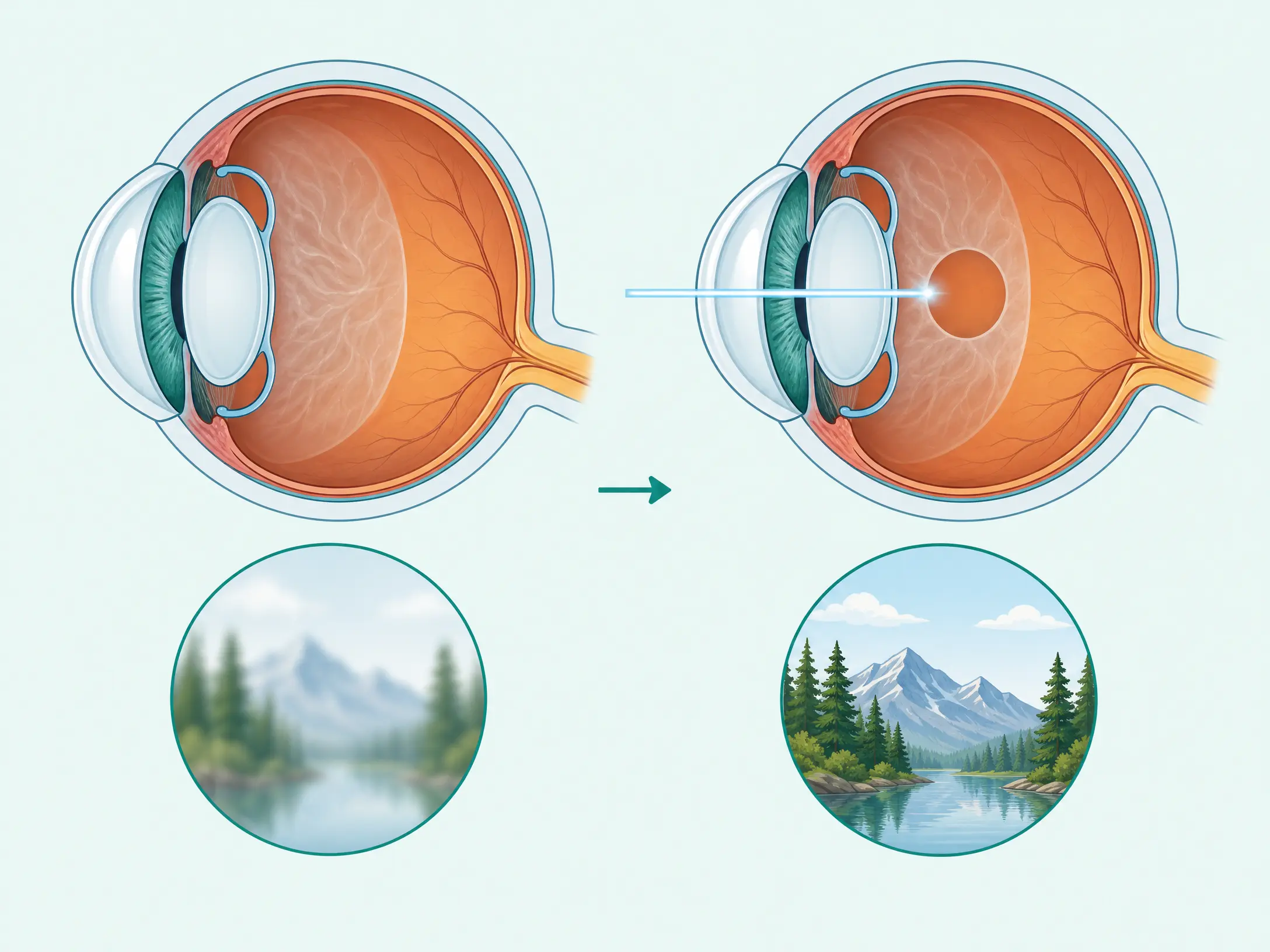
A misting of the thin membrane behind an artificial lens, months or years after cataract surgery. It is not the cataract returning, and it clears in minutes with a YAG laser.

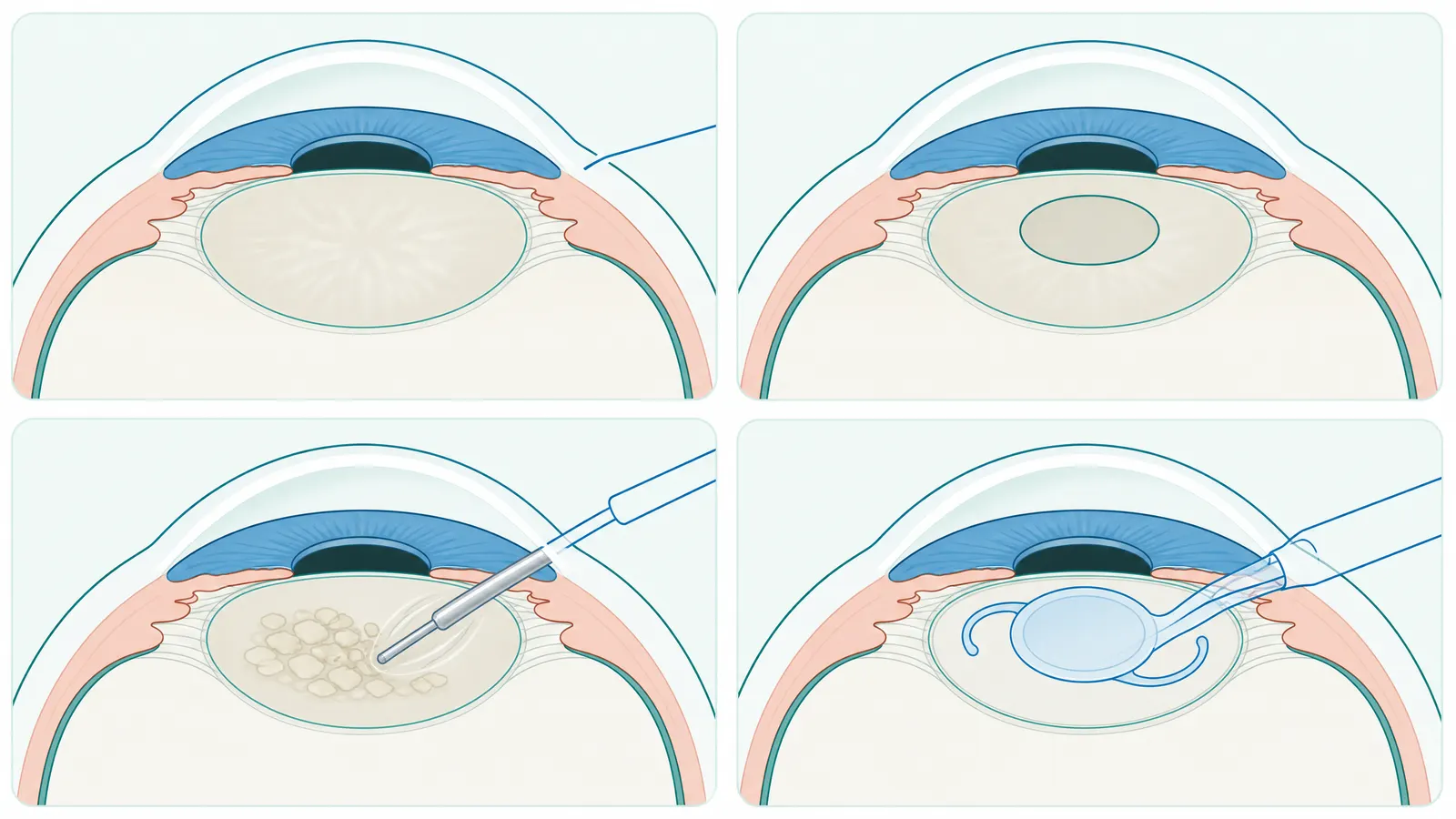
Four-step diagram of phacoemulsification cataract surgery with IOL implantation

Phacoemulsification Phaco

Procedure

The standard cataract operation: through a tiny keyhole, ultrasound breaks up the cloudy lens, it’s removed, and a folded artificial lens unfolds into place — usually stitchless daycare.

See also Cataract IOL FLACS

Phakic IOL

Procedure

A corrective lens implanted inside the eye while keeping your own natural lens — the category the ICL belongs to. Used for stronger prescriptions.

See also ICL IOL
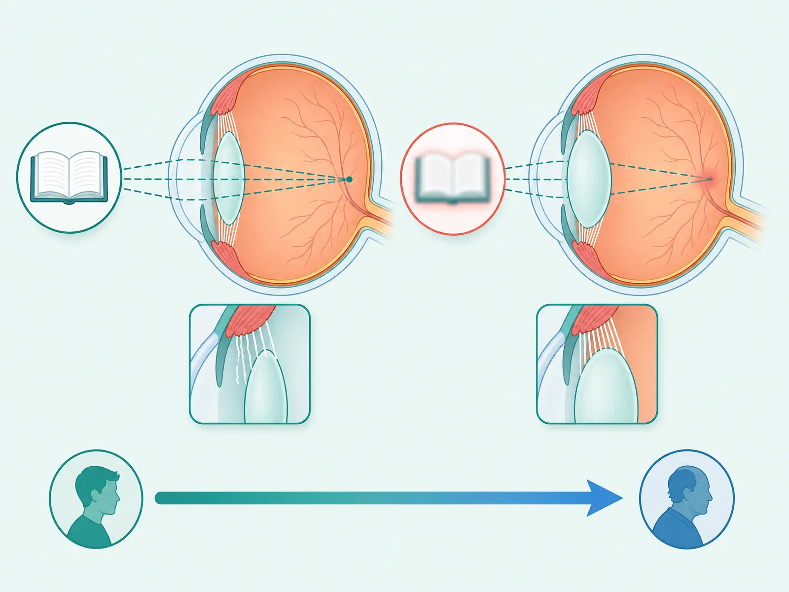
Diagram explaining age-related loss of near focus (presbyopia)

Presbyopia

Condition

The natural stiffening of the lens after about age 40, so near focus fades and reading drifts to arm’s length. Everyone experiences it eventually.

PRK Photorefractive keratectomy

Procedure

Laser vision correction that reshapes the corneal surface directly, with no flap. A good choice for thinner corneas or active lifestyles; recovery is a little slower than LASIK.

See also LASIK SMILE Cornea

Pupil

Anatomy

The black opening at the centre of the iris that lets light into the eye — widening in dim light and narrowing in bright.

See also Iris

PVD Posterior vitreous detachment

Condition

The vitreous gel separating from the retina with age. Common and usually harmless, but new flashes or a burst of floaters should be checked, as it can occasionally tear the retina.

R
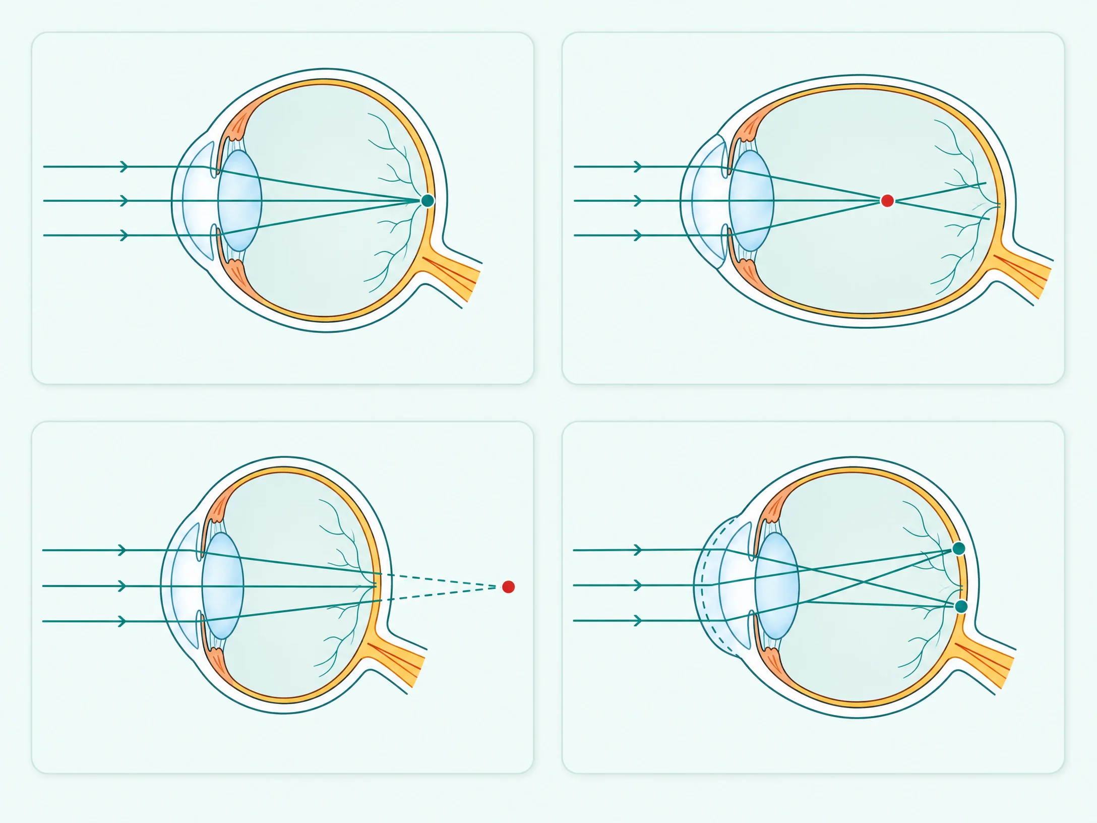
Diagram comparing normal focus, myopia, hyperopia and astigmatism

Refractive Error

Condition

The umbrella term for myopia, hyperopia and astigmatism — where the eye doesn’t focus light precisely on the retina. Corrected with glasses, contact lenses or laser surgery.

Retina

Anatomy

The light-sensitive lining at the back of the eye that captures images and sends them to the brain — like the film in a camera.

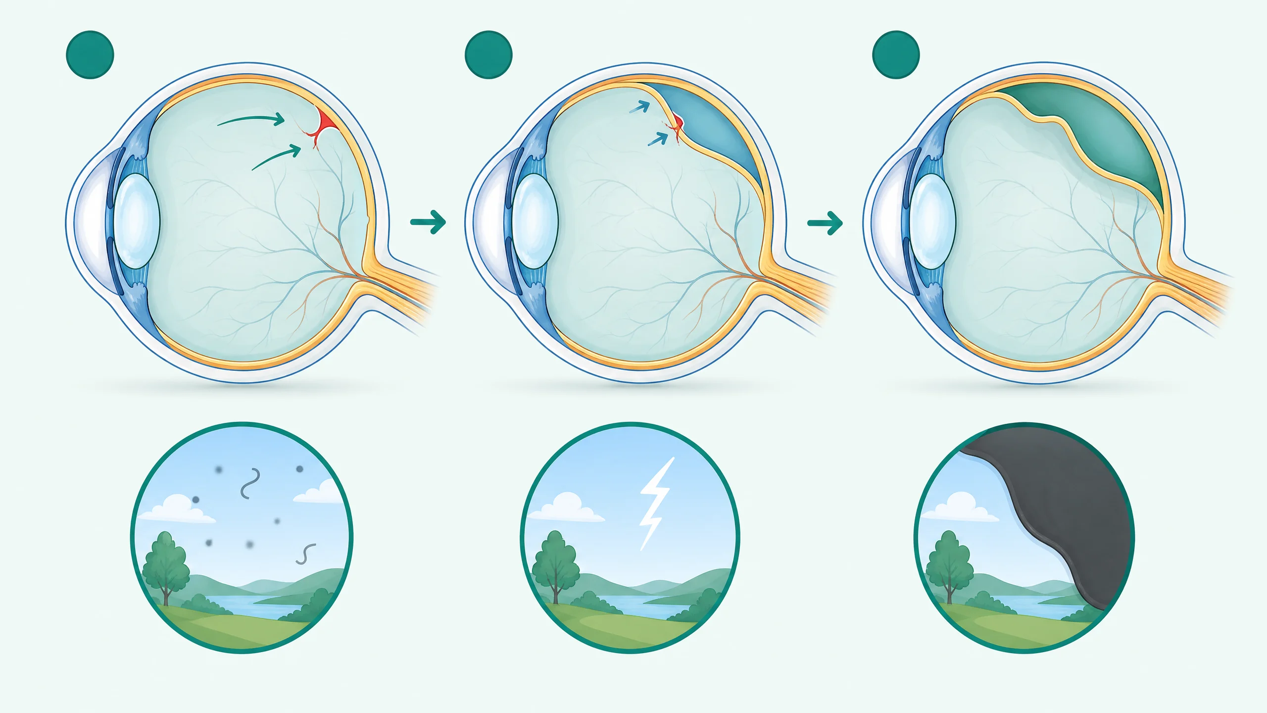
Diagram of retinal tear progressing to retinal detachment with warning signs

Retinal Detachment

Condition

A sight-threatening emergency where the retina peels away from the back wall of the eye. Warning signs are new floaters, flashes and a dark curtain over vision — seek help the same day.

See also Retina PVD Floaters
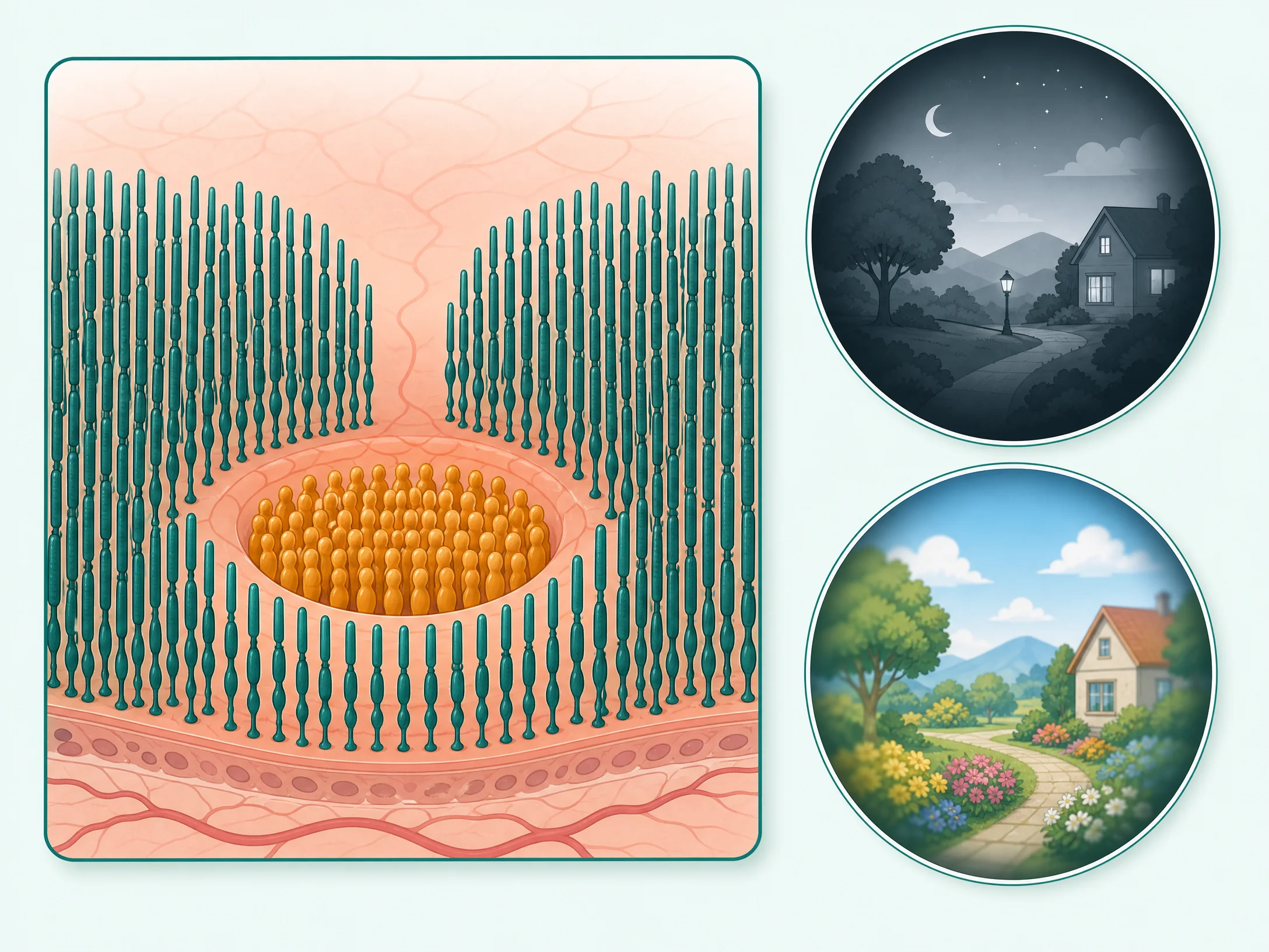
Diagram of rod and cone photoreceptors and their roles

Rods & Cones

Anatomy

The retina’s two photoreceptor cells. Rods (~120 million) handle dim light and side vision; cones (~6 million, packed in the macula) handle colour and fine detail.

See also Retina Macula
S

Sclera

Anatomy

The tough white outer coat of the eye that protects it and holds its shape.

See also Cornea

SMILE Small-incision lenticule extraction

Procedure

Flapless laser vision correction: a laser shapes a thin lens of tissue inside the cornea and removes it through a keyhole 2–4 mm opening. Minimal disturbance to the surface.

See also LASIK PRK Cornea
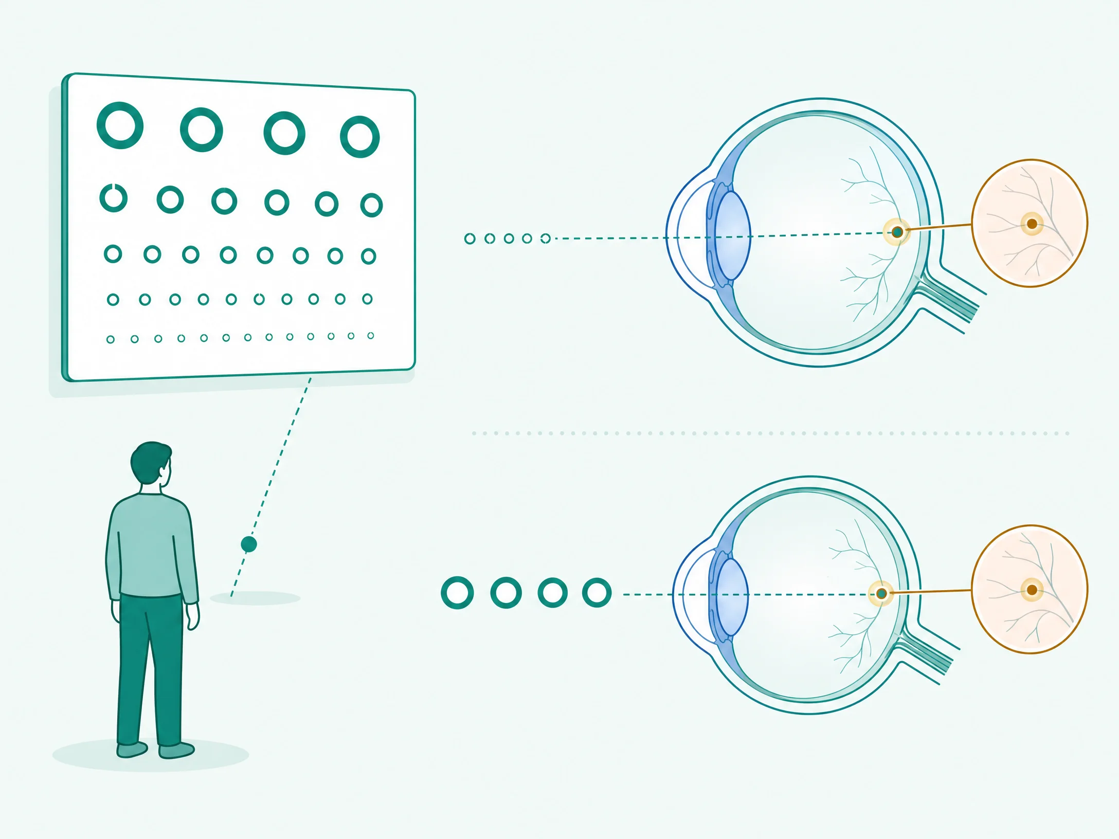
Diagram explaining how visual acuity is measured with a distance chart

Snellen Chart

Test

The classic wall chart of shrinking letters used to measure distance vision. Reading the "6/6" line means normal distance clarity.

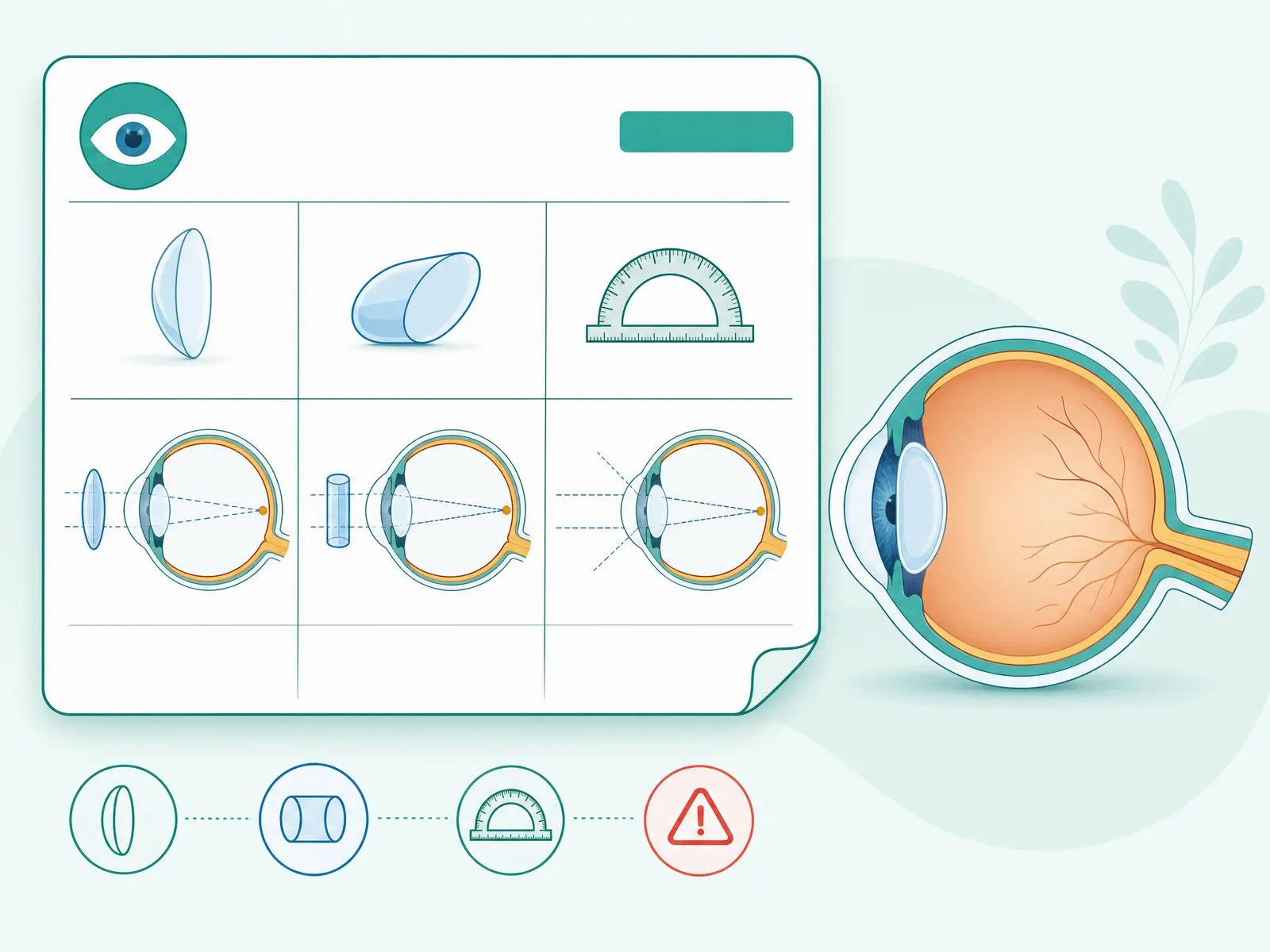
Diagram explaining the SPH, CYL and AXIS values on a glasses prescription

Spherical Power SPH

Optics

The main power on your glasses prescription — a minus value corrects myopia, a plus value corrects hyperopia.

Diagram of different types of eye misalignment (squint)

Squint Strabismus

Condition

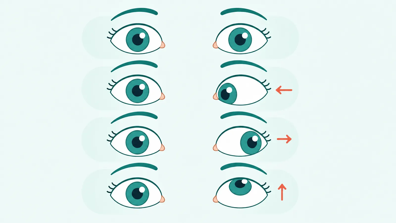
When the two eyes don’t point in the same direction — one may turn in, out, up or down. Treatable at any age, and important to catch early in children.

See also Amblyopia
T
Diagram of the three tear film layers and an unstable tear film in dry eye

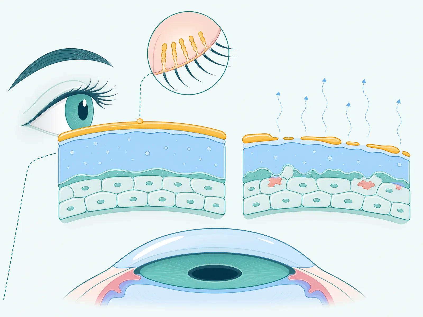
Tear Film

Anatomy

The thin three-layer coating (oil, water, mucin) that keeps the eye’s surface smooth and comfortable. When it breaks down, dry eye follows.

See also Dry Eye

Toric

Optics

An artificial lens (or contact lens) built to correct astigmatism, with orientation marks so it sits at the right angle.

See also IOL Astigmatism

Trabecular Meshwork

Anatomy

The tiny drainage filter at the angle where the iris meets the cornea. When it drains poorly, pressure builds — the mechanism behind most glaucoma.

See also Glaucoma Iris
V

Visual Acuity

Test

A measure of how clearly you see fine detail at distance, recorded as values like 6/6 and tested on a Snellen chart.

Vitreous Vitreous humour

Anatomy

The clear gel that fills the large cavity between the lens and the retina, giving the eyeball its shape. It shrinks with age, causing floaters.

See also Floaters PVD Retina
Y
Diagram of posterior capsule opacification cleared by YAG laser capsulotomy

YAG Capsulotomy

Procedure

A quick, painless clinic laser that opens a clear window in the misted membrane behind an artificial lens (PCO), restoring sharp vision in minutes.

See also PCO IOL

Illustration and definitions simplified for education — they don't replace an eye examination. For a diagnosis specific to you, book a consultation with our doctors.

Still Have a Question?

A term on your report you don't recognise?

Bring it in — our medical counsellors will explain exactly what it means for your eyes. No Sales, Expert Consultation.

+91 81786 39701
Book an Appointment

Medically reviewed by Dr. Sanjeev Bisla, MBBS, MS (Ophthalmology), Director — Dayal Eye Centre · Last reviewed: 24 July 2026 · Editorial Policy