LASIK surgery can treat myopia effectively, but studies show regression happens more often than most people think. Research reveals that patients experienced an average regression of -1.66 diopters over a 15-year period. The yearly regression rate stands at -0.11 diopters.
LASIK delivers excellent results for myopia treatment right after surgery. The data shows that after 10 years, myopia greater than 1 diopter developed in 67% of treated eyes. Scientists haven’t fully understood this regression yet. The process has multiple factors that range from corneal healing to inflammatory responses.
This piece will help you understand what triggers LASIK regression. We’ll get into the risk factors and show you the long-term results of this popular vision correction procedure.
Understanding LASIK and Vision Changes
The cornea serves a vital role in vision. It accounts for about 80% of the eye’s total refractive power. During LASIK surgery, a precise laser reshapes this transparent tissue. This corrects refractive errors, especially when you have myopia where light rays focus incorrectly in front of the retina.
How LASIK corrects myopia
LASIK creates a hinged lamellar corneal flap. An excimer laser makes calculated adjustments to the stromal bed underneath. Your cornea has five distinct layers. The stroma makes up 90% of its thickness. This stromal layer contains about 200-250 lamellae – flattened bundles of collagen that stretch across the cornea’s width.
Surgeons must carefully measure corneal tissue before removal. These measurements need repeating at least a week before surgery to ensure stability. On top of that, the surgical plan looks at factors like optical zone diameter and transition zones. These elements substantially affect long-term outcomes.
Normal healing vs regression
After LASIK, most patients see at 20/20 or better. Yet some people experience regression – their vision gradually returns toward their original prescription. Studies show less than 5% of patients notice regression in the first year.
Biology can trigger regression in several ways. Your cornea might gradually change back toward its original shape. This happens more often in patients with thinner corneas or those who need correction for high degrees of myopia. Changes in hormones during pregnancy or menopause can also affect corneal thickness and possibly influence post-LASIK stability.
Timeline of vision changes
LASIK recovery follows a predictable pattern. Many patients see better right away, though temporary blurriness occurs during the first phase of recovery. The first 24 hours might bring burning sensations, excessive tearing, or a gritty feeling in your eyes.
Vision typically stabilizes within 2-3 months after surgery. During this time, patients might experience:
- Fluctuating vision clarity
- Changes in night vision
- Occasional dry eye symptoms
Complete healing takes up to six months as your cornea adapts to its new shape. Eye doctors schedule regular follow-up appointments throughout this period. This helps them monitor healing progress and address concerns quickly.
Success rates vary based on your original prescription strength. Patients with mild nearsightedness usually get more predictable results than those with severe myopia or combined astigmatism. The type of laser system affects long-term stability too – flying spot machines show lower regression risks than scanning slit systems.
Early Signs of LASIK Regression
Patients need to watch for subtle vision quality changes to spot LASIK regression. Research shows that regression happens slowly as patients’ vision partially returns to their pre-surgery state.
Gradual blur development
Vision correction after LASIK tends to deteriorate slowly. Research indicates that myopic regression advances at -0.11 diopters yearly. Patients might experience an average regression of -1.66 ± 2.15 diopters over 15 years.
The largest longitudinal study reveals troubling regression statistics. All but one of these patients developed myopia exceeding 1 diopter after 10 years. These changes often start early, and patients notice differences within their first post-surgery year.
Blurred vision can develop due to several factors:
- Corneal healing responses
- Fluctuating blood sugar levels
- Extended screen time exposure
- Environmental factors
- Underlying health conditions
Dry eye symptoms can look like regression and cause temporary vision fluctuations that get better with blinking. Vision clarity becomes unstable when dryness persists without treatment.
Changes in night vision
Night vision quality changes rank among the most important signs of LASIK regression. Patients often report increased light sensitivity and visual problems in low-light settings.
Night vision challenges show up as:
- Halos around bright lights
- Increased glare sensitivity
- Starburst effects
- Reduced contrast sensitivity
- Double vision in dim conditions
Patients may struggle with low-light vision quality even when standard tests look good. The halo distortion index usually jumps by 2.15 after LASIK, whatever the procedure’s success by normal standards.
These night vision issues affect about 43.5% of patients who get refractive surgical procedures. Modern surgical techniques haven’t eliminated spherical aberration and secondary astigmatism changes that lead to these visual problems.
Vision changes can last beyond the normal recovery time for patients with regression symptoms. Signs of regression might appear 1 to 3 months after surgery and worsen over several years. Early regression signs often link to corneal thickness changes and shifts in corneal curvature.
Doctors track these changes through regular checkups. They might suggest more treatments if regression symptoms continue or get worse. About 4% of patients with early myopic progression after SMILE surgery and 20% after LASIK need additional procedures to fix their vision.
Biological Causes of Regression
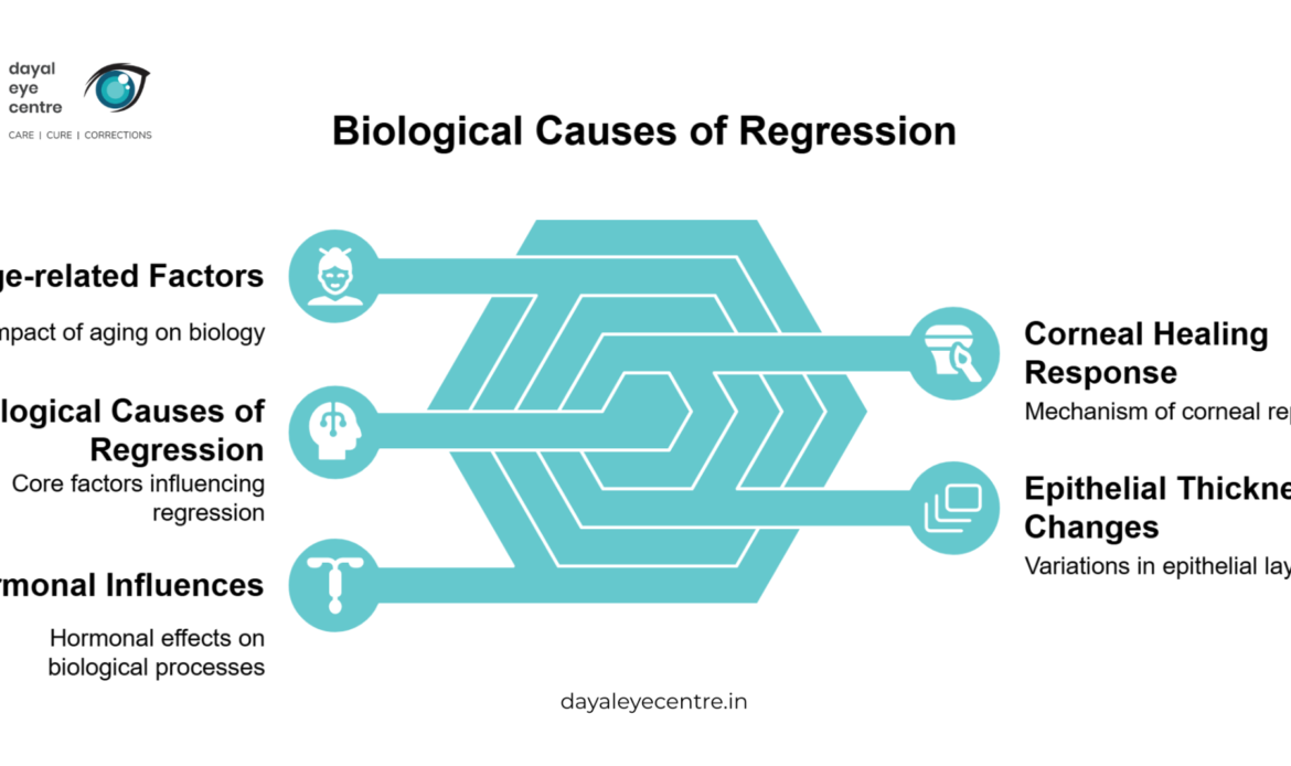
Several biological mechanisms cause vision regression after LASIK surgery. These range from cellular responses to age-related changes. Doctors can better explain why some patients gradually return to their original prescription by knowing these factors.
Corneal healing response
Complex cellular interactions trigger the corneal healing process right after surgery. Research shows that keratocyte activation is crucial, and the thickness of the keratocyte activation zone reaches 21.54 μm one week after surgery. This activation zone then decreases to 8.75 μm after one month and becomes almost undetectable six months later.
Cytokine-mediated wound healing cascades help replace lost corneal cells. The prolonged elevation of cytokine signaling often produces more cells than needed for surface integrity, which ended up causing regression.
Epithelial thickness changes
LASIK surgery causes the corneal epithelium to reshape extensively. Scientists have found that complete epithelial turnover happens every 5-7 days. The epithelium can quickly renew itself to fix stromal irregularities, but this ability also leads to regression.
Studies show epithelial thickening varies in different corneal areas:
- Central thickening averages 4μm
- Mid-peripheral thickening reaches about 7μm
The largest epithelial response happens within the first month. This response matches a -0.39 D move in refraction. Such thickening creates a negative meniscus-like effect that might undo the original surgical correction.
Age-related factors
Patient age affects LASIK outcomes by a lot. Research shows older patients (>55 years) are as safe as younger patients (30-40 years) during surgery but get less effective results. This happens because:
- Older patients heal more slowly
- Corneal biomechanical properties change
- Stromal collagen framework undergoes structural changes
Hormonal influences
Hormones greatly impact post-LASIK stability. Women on hormone replacement therapy have a higher chance of refractive regression. The risk of regression after photorefractive procedures is 13.5 times higher in women taking oral contraceptives.
Estrogen affects corneal stability through several ways:
- Gets matrix metalloproteinases to produce more
- Makes collagenases active
- Produces more prostaglandin
- Breaks down type I collagen
Pregnancy increases estrogen levels about twenty times, though progesterone helps counter its biomechanical effects. These hormone changes explain why pregnant women often can’t tolerate contact lenses and why their post-LASIK vision might change during pregnancy.
Risk Factors That Increase Regression
Research shows that specific patient traits and anatomical features affect the chances of LASIK regression. Doctors can better predict vision changes after surgery by knowing these risk factors.
High original prescription
Higher degrees of myopia lead to increased regression rates. Patients with moderate to high myopia show an average regression of -1.66 diopters over 15 years. This means they experience about -0.11 diopters of regression each year.
Regression rates change based on myopia severity:
- Low to moderate myopia: 10% regression rate
- High myopia: 30% regression rate
Data analysis shows a clear link between pre-operative spherical equivalent and myopic regression (correlation coefficient: 0.333). This higher risk comes from:
- More corneal tissue removal needed for higher corrections
- Higher biomechanical stress on remaining corneal tissue
- Bigger changes to corneal shape
Thin corneas
Corneal thickness plays a key role in LASIK results. Patients with corneas thinner than 500 micrometers face special challenges. Young patients’ thin corneas tend to show more refractive regression.
Research has looked at how corneal thickness relates to regression:
- Corneas thinner than 500 μm before surgery associate with higher regression rates
- Residual stromal bed thickness affects long-term stability
- Regression groups show bigger differences between apex and thinnest corneal points after surgery
Recent findings have changed what we know about thin corneas. LASIK can work safely in corneas under 500 μm when other risk factors aren’t present. Doctors need to look at several factors:
- Pre-operative corneal curvature measurements
- Planned ablation depth
- Residual stromal bed thickness
- Overall corneal biomechanics
Corneal thickness and regression have a more complex relationship than we once thought. Similar pachymetry values don’t always mean corneas have the same strength. The mix of thickness, biomechanical properties, and other anatomical features determines regression risk.
Modern surgery includes preventive steps for high-risk cases. LASIK with corneal collagen cross-linking (LASIK Xtra) helps reduce regression rates. This combined approach makes corneal tissue stronger and might give better long-term results for patients with risk factors.
How Doctors Diagnose Regression
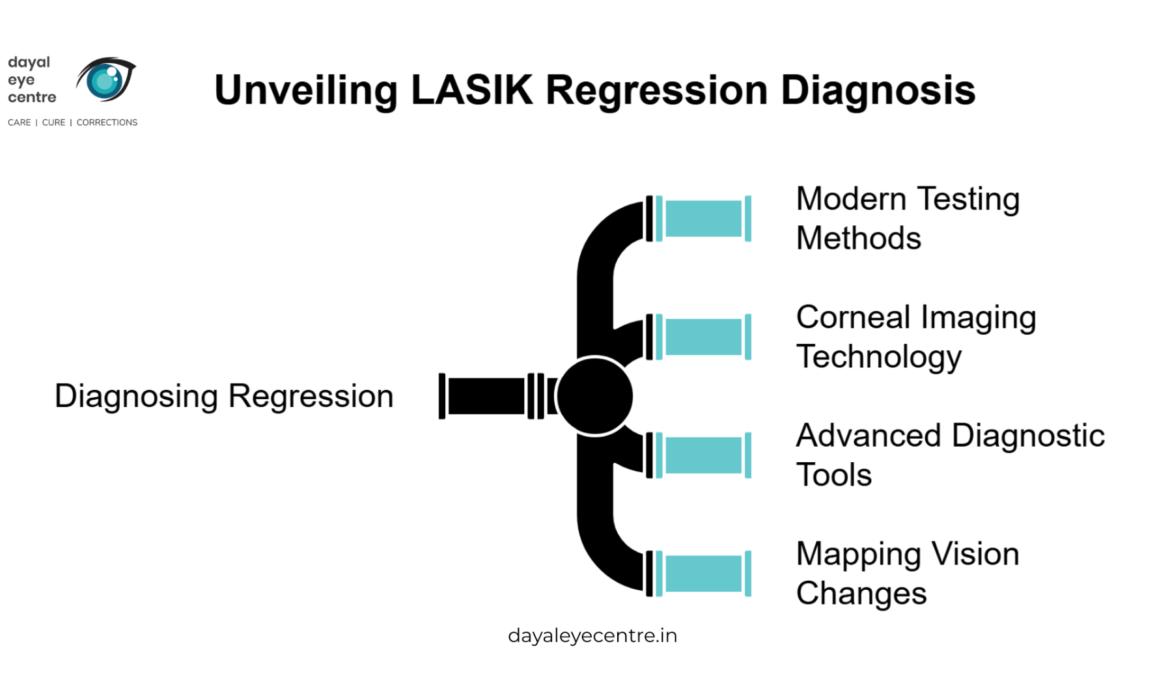
Medical professionals need sophisticated equipment and complete testing protocols to diagnose regression after LASIK. Eye doctors use advanced techniques that detect even small changes in vision quality and corneal structure.
Modern testing methods
Doctors use multiple diagnostic approaches to identify regression. A detailed evaluation has cycloplegic refraction, which showed measurements of -1.25 DS in right eyes and -3.75 DS in left eyes among regression cases. Standard vision tests and corneal interface examinations often reveal slight haziness in regression cases.
Medical professionals have different definitions of regression, but most think about a change of 0.25 diopters or greater as clinically important. They analyze:
- Manifest spherical equivalent
- Cycloplegic measurements
- Central corneal curvature
- Intraocular pressure
- Schirmer test results
Mapping vision changes
Doctors track vision changes through normalized topography and compare current maps with pre-operative readings. New advances in corneal mapping show distinctive patterns, including peripheral hot rings that point to regression.
Advanced diagnostic tools measure several vital parameters:
- Ablation depth measurements
- Optical zone diameter assessment
- Flap thickness evaluation
- Residual stromal bed thickness
Research shows flap thickness typically measures around 110.00 ± 0.00 μm in FS-LASIK procedures. The ablation depth averages 104.82 ± 29.64 μm, while lenticular thickness measures 133.24 ± 25.74 μm.
Corneal imaging technology
Optical coherence tomography (OCT) is the life-blood of regression diagnosis. This technology helps doctors examine epithelial mapping without compensatory central hyperplasia. Doctors also use Pentacam Scheimpflug densitometry that detects subtle interface scarring through measurements ranging from 25.4 to 28.2 Gray Scale Units.
Advanced imaging systems give several benefits:
- Precise measurement of corneal layers
- Detection of early regression signs
- Assessment of healing patterns
- Evaluation of biomechanical changes
Doctors now rely on wavefront analysis to spot regression and corneal irregularities. This technology helps detect higher-order aberrations that might go unnoticed through regular testing methods.
The Cox proportional hazard model helps evaluate myopic regression. Studies show cumulative survival rates at 12 and 18 months after surgery reach 55.07% and 43.03% respectively for FS-LASIK procedures.
Light propagation algorithm analysis marks another breakthrough in regression diagnosis. This method calculates corneal transmittance using height data from both corneal surfaces with local thickness measurements. The technique gives a complete analysis of the whole corneal surface instead of just using average keratometry values.
Conclusion
Knowing how LASIK changes over time helps patients make better decisions about their vision correction experience. Studies show that LASIK works well to treat myopia, but vision changes happen gradually. Research suggests an average change of -1.66 diopters over 15 years.
Your original prescription strength and corneal thickness are the foundations of how your vision might change. Your body’s healing response and hormone levels are also important factors. Eye doctors need to screen patients carefully to achieve the best results.
Eye care professionals now use sophisticated tools like OCT and wavefront analysis to spot early warning signs. These tools help doctors track small changes in vision and fix problems quickly. Regular visits to experienced eye specialists help catch and manage any vision changes after LASIK.
LASIK delivers great results right away. Patients should know their vision might change somewhat over time. Each person’s results depend on their unique factors. That’s why customized treatment plans and regular monitoring are vital parts of lasting vision correction.
FAQs
1. How common is regression after LASIK surgery?
Studies show that regression after LASIK is more common than many realize. Over a 15-year period, patients experienced an average regression of -1.66 diopters, with a yearly regression rate of -0.11 diopters. After 10 years, about 67% of treated eyes developed myopia greater than 1 diopter.
2. What are the early signs of LASIK regression? Early signs of LASIK regression include gradual blur development, changes in night vision such as increased light sensitivity and visual disturbances under low-light conditions, and fluctuations in vision clarity. These changes often begin within the first year following surgery.
3. What biological factors contribute to LASIK regression?
Biological factors contributing to LASIK regression include corneal healing responses, epithelial thickness changes, age-related factors, and hormonal influences. The corneal epithelium’s rapid renewal capability and hormonal fluctuations, particularly in women, can significantly impact post-LASIK stability.
4. Are certain individuals more at risk for LASIK regression?
Yes, individuals with high initial prescriptions and thin corneas are at higher risk for LASIK regression. Patients with moderate to high myopia and those with corneas thinner than 500 micrometers face increased chances of experiencing regression after surgery.
5. How do doctors diagnose and monitor LASIK regression?
Doctors use advanced diagnostic tools to diagnose and monitor LASIK regression, including cycloplegic refraction, corneal topography mapping, optical coherence tomography (OCT), and wavefront analysis. These technologies allow for precise measurement of corneal layers and detection of early regression signs.