More than 16 million people worldwide have transformed their lives through refractive surgery procedures. The success rate stands at an impressive 98% for achieving perfect 20/20 vision. These procedures now take just 30 minutes and help 8 out of 10 patients live without glasses or contact lenses.
LASIK surgery leads the way as the preferred choice for vision correction, though patients can choose from several refractive surgery options. The progress of these procedures traces back over a century, from theoretical foundations in 1885 to today’s sophisticated laser-based techniques.
This detailed piece explains how different refractive surgery procedures work. You’ll learn everything from the original consultation to the final cornea reshaping steps. We’ll also look at the state-of-the-art technology that drives the success of these vision-correcting procedures.
The Evolution of Refractive Surgery
The story of refractive surgery started in 1885 at the time Norwegian ophthalmologist Hjalmar August Schiøtz published his first theoretical work about its potential. Japanese ophthalmologist Tsutomu Sato took the next step in 1930 and made groundbreaking attempts by performing radial cuts in the cornea to correct vision by up to 6 diopters.
Early surgical techniques
Jose Barraquer made a breakthrough in 1963 at his ophthalmologic clinic in Bogotá, Colombia. His revolutionary technique, keratomileusis, could correct both myopia and hyperopia. The process involved removing a corneal layer, freezing it for manual sculpting, and putting the reshaped layer back into the eye.
Svyatoslav Fyodorov then introduced Radial Keratotomy (RK) in the USSR in 1974. His method used precise diamond knife incisions in the cornea to change its shape.
Introduction of laser technology
Refractive surgery went through a dramatic change with the creation of the excimer laser. Scientists first experimented with xenon dimer in 1970 and noble gas halides in 1975. IBM scientist Rangaswamy Srinivasan found in 1980 that the excimer laser could cut organic tissues with incredible precision without causing major thermal damage.
Stephen Trokel worked with Theo Seiler and Srinivasan to perform the first Photorefractive Keratectomy (PRK) in Germany in 1983. This procedure, first known as keratomileusis in situ, launched the era of laser-based refractive surgery.
Modern innovations
LASIK (Laser-Assisted In Situ Keratomileusis) came next, patented by Gholam Ali Peyman in 1989. This breakthrough combined creating a corneal flap with excimer laser reshaping, which helped patients recover faster than earlier techniques.
The 1990s brought major technological breakthroughs. J.T. Lin got a US patent for flying-spot technology for customized LASIK in 1991, which surgeons use worldwide today. Dr. S. Lai created eye-tracking technology in 1993 to prevent decentralisation during LASIK procedures.
Femtosecond laser technology arrived in 2001. This technology eliminated the need for mechanical blades in flap creation and made the procedure safer and more precise. Wavefront-guided and topography-guided systems have refined these procedures even further. They now treat higher-order aberrations and provide tailored vision correction.
Small Incision Lenticule Extraction (SMILE) has emerged as a minimally invasive option that works especially well for treating myopia. The procedure uses femtosecond laser technology without needing a corneal flap, marking the latest achievement in refractive surgery’s progress.
Understanding Different Refractive Procedures
Modern refractive surgery gives you three main procedures that work for different patient needs. Understanding these procedures helps you make better decisions about vision correction options.
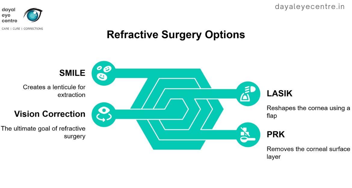
LASIK surgery process
LASIK is the most common laser vision surgery that works well to treat myopia, hyperopia, and astigmatism. The surgeon creates a corneal flap with a femtosecond laser or mechanical microkeratome. They fold back the flap and sculpt the underlying stromal bed with an excimer laser. The flap goes back in place without needing sutures.
LASIK’s biggest advantage is quick visual recovery – patients see better by the next day. But complications can happen with the flap like irregular formation, dislocation, and long-term corneal ectasia when the cornea gets too thin and unstable.
PRK procedure steps
PRK is different from LASIK because it doesn’t need a corneal flap creation. The surgeon removes the corneal epithelium first and then uses an excimer laser to sculpt the anterior corneal stromal bed. The epithelium grows back in 3-4 days while patients wear a bandage contact lens.
PRK works great if you have thin corneas or epithelial basement membrane dystrophy. The procedure leaves a thicker residual stromal bed which lowers ectasia risk. Patients might see corneal haze, especially when substantial corneal tissue needs ablation. Recovery takes 3-4 days, and most patients feel comfortable with proper medication.
SMILE technique overview
SMILE shows the newest advances in refractive surgery. The surgeon uses a femtosecond laser to create a thin, intrastromal lenticule and takes it out through a small 2-4mm peripheral corneal incision. This procedure treats myopia and myopic astigmatism as well as LASIK does.
SMILE’s best feature is its minimal invasion – it needs just a tiny cut instead of a full corneal flap. This leads to less corneal denervation and quicker nerve healing than LASIK. The challenges include higher risk of suction loss during surgery and tougher enhancement surgeries.
SMILE takes about 20 minutes under local anesthesia. Patients see better within 24 hours, though full recovery needs several days. Studies show that 99% of patients achieve at least 20/40 vision within six months, and 88% get to 20/20 vision.
Pre-Surgery Assessment and Planning
A full pre-operative assessment is the life-blood of successful refractive surgery. This vital phase helps doctors pick the best treatment approach and predict how well the surgery might work.
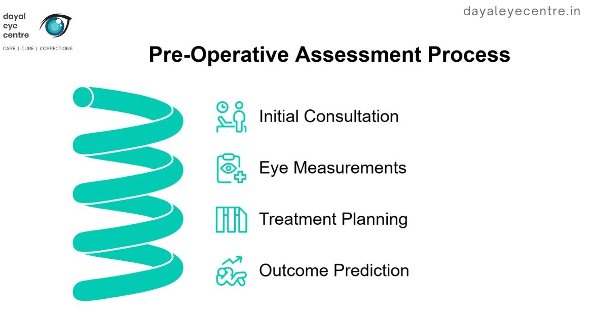
Original consultation
Your consultation usually takes 30-45 minutes and gives a detailed picture of your eye health and vision needs. Patients need to stop wearing contact lenses before their appointment – one week for soft non-toric lenses, two weeks for toric lenses, and at least three weeks for rigid lenses. This break will give accurate corneal measurements because contact lenses can change the cornea’s physical properties.
During the consultation, doctors run several key tests:
- Tonometry to measure intraocular pressure
- Visual acuity assessment
- Detailed examination of anterior and posterior eye segments
- Full corneal analysis including topography and pachymetry
Your eye doctor will review your medical history and ask about medications or supplements that might affect your surgery results. They also assess conditions like diabetes that could impact how well you heal.
Eye measurements and mapping
Precise corneal measurements are the foundation of pre-surgery planning. Eye doctors employ advanced diagnostic tools to map your cornea’s shape, thickness, and structure.
Corneal topography is a basic tool that creates three-dimensional maps and shows changes across the corneal surface. This technology helps doctors:
- Spot early signs of conditions like keratoconus
- Plan custom treatment approaches
- Get corneal thickness profiles
Wavefront technology gets unique imperfections in your cornea that let surgeons customize the procedure for your eyes. The IOL Master measures axial eye length with better precision than traditional techniques.
Without doubt, pupil measurement is vital in pre-operative assessment. Doctors use various tools, like the Colvard Pupillometer or Procyon device, to measure pupil size in different lighting conditions. These measurements help avoid problems with night vision after surgery.
The detailed evaluation looks at corneal curvature since post-refractive ectasia remains a big concern. Eye doctors carefully check several risk factors:
- Abnormal topography readings
- Percentage of tissue alteration
- Corneal thickness measurements
- Age considerations
- Degree of myopia
Before setting the final surgical plan, doctors use AI-based systems like the Pentacam Random Forest Index (PRFI) that has shown great accuracy in finding mild ectasia. The tomographic and biomechanical index (TBI) also provides 90.4% sensitivity and 96.0% specificity to identify subclinical ectasia.
Step-by-Step Surgery Process
Refractive surgery procedures follow a careful sequence that ensures the best outcomes through exact surgical steps. Let’s look at how ophthalmologists reshape your cornea and fix vision problems.
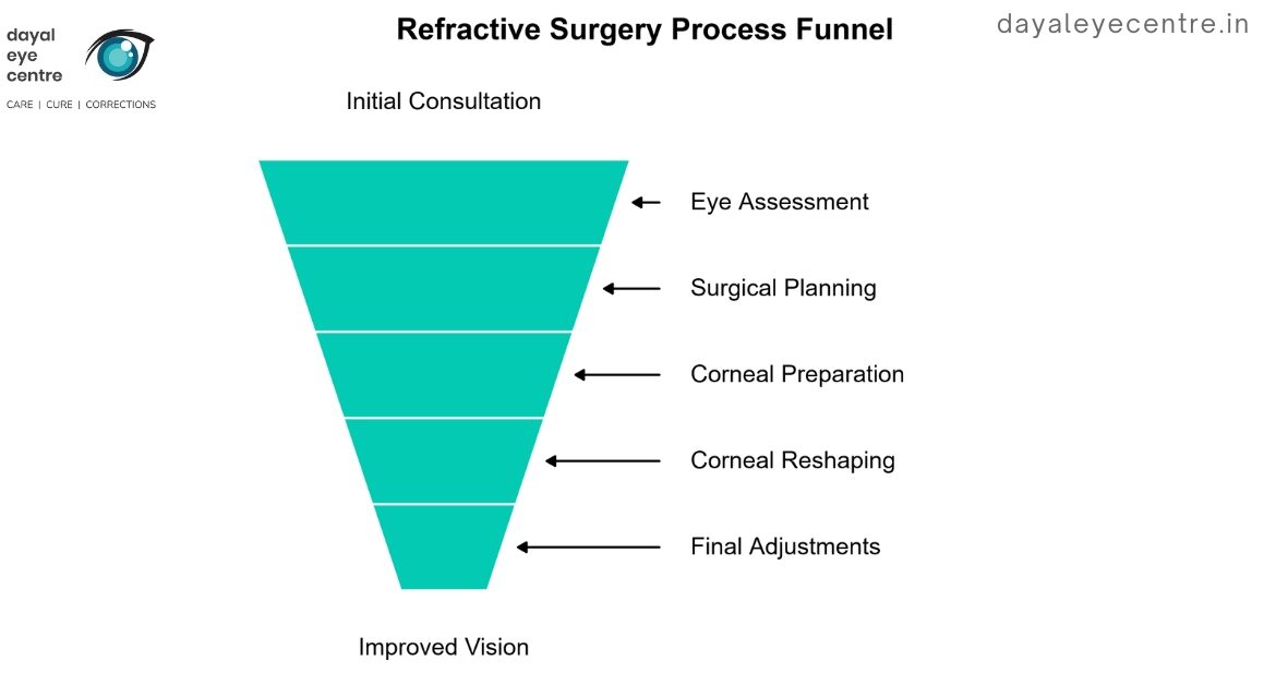
Preparing the eye
The surgery starts when your doctor applies numbing eye drops to keep you comfortable during the procedure. Your surgeon uses a lid speculum, a special tool that keeps your eyelids open. This step is vital because it stops you from blinking and gives clear access to your eye.
The surgeon then marks key points on your cornea and places you under the laser system. You’ll need to look at a specific light point that helps keep your eye lined up properly.
Creating corneal access
Your surgeon will access your cornea based on the procedure you choose. LASIK surgery needs a thin corneal flap (100-200 micrometers) made with either a femtosecond laser or mechanical microkeratome. This flap works like a hinged door to reach the stromal tissue underneath.
PRK works differently. The surgeon removes the epithelium – your cornea’s outer layer – using mechanical, chemical, or laser methods. This gives direct access to the stromal bed without making a flap.
Reshaping the cornea
The reshaping uses advanced laser technology, mainly the excimer laser operating at 193 nanometers. The laser removes a round piece of tissue from your cornea’s center to fix nearsightedness by flattening it. For farsightedness, the laser treats the cornea’s outer edges to increase its central curve.
Advanced eye-tracking systems watch and adjust for any eye movements to ensure exact tissue removal. The laser sends carefully measured pulses based on your pre-operative measurements, and each pulse removes tiny amounts of tissue.
Final adjustments
Your surgeon makes final touches based on live assessment. In LASIK, they carefully put the corneal flap back in place. The flap sticks naturally without stitches because of your endothelium’s pumping effect. Tissue growth makes the flap permanent within hours.
PRK patients get a bandage contact lens to protect the treated area and help epithelial healing. This lens stays in place for several days while the epithelium grows back.
The whole surgery usually takes less than 30 minutes. Your surgeon checks to make sure the flap is in the right position or the epithelium is properly covered right after the procedure. Most people see better within 24 hours, but full stability might take several weeks.
Technology Behind Modern Procedures
Advanced technology serves as the foundation of modern refractive surgery. It makes unprecedented precision and safety possible in vision correction procedures. Let’s take a closer look at the sophisticated systems behind these procedures.
Laser systems
The excimer laser operates at 193 nanometers and is the life-blood of refractive procedures. This remarkable system combines reactive gasses like chlorine and fluorine with inert gasses such as argon, krypton, and xenon. These gasses form an excited-state compound when they receive a high-voltage electrical discharge. The compound releases precise energy pulses that reshape the cornea.
The excimer laser’s 193nm wavelength works perfectly because it reaches only the superficial corneal layer (0.3 μm). Such precision affects surrounding tissues minimally, as each pulse removes about 1-μm of corneal tissue at 1-J/cm2 energy density.
Today’s laser systems have evolved beyond traditional full-beam delivery. They now include:
- Scanning beams with smaller spot sizes (0.6 to 2.0 mm)
- Flying spot technology for improved precision
- Sophisticated ablation algorithms for smoother transitions
- Advanced cooling mechanisms to protect surrounding tissue
The femtosecond laser brings another breakthrough by creating precise corneal flaps without mechanical blades. This ultrafast laser delivers unique accuracy that improves surgical safety and overall outcomes.
Eye tracking technology
Sophisticated eye-tracking systems mark a crucial advancement in refractive surgery. These systems track eye movements in multiple dimensions:
- Lateral movements (up and down)
- Horizontal and vertical movements from head tilts
- Rotational movements around the optical axis
- Z-level adjustments for upward or downward eye movements
Modern tracking systems can detect movements up to 4,000 times per second. This exceptional speed allows up-to-the-minute laser adjustments that ensure precise alignment throughout the procedure. The system automatically pauses the laser if excessive eye movement occurs, which prevents potential complications.
Advanced Eye-Tracking Systems
The Advanced Control Eye-tracking (ACE) system shows this technology’s sophistication. It creates a detailed iris map before surgery to establish a foundation for multi-dimensional tracking. This approach ensures that treatment profiles match the correct patient and eye, which eliminates wrong-eye procedure concerns.
Artificial intelligence has improved these systems by enabling smart analysis of patient data and more precise treatment planning. These AI-powered platforms help achieve higher success rates and better patient satisfaction by optimizing diagnostic accuracy and treatment outcomes.
Modern eye-tracking systems work at remarkable speeds and communicate directly with the laser to ensure unique precision. The laser repositions instantly with even the slightest eye movement to maintain perfect alignment throughout the procedure. On top of that, integrated sensors monitor head position to provide extra safety during surgery.
Conclusion
Refractive surgery has changed from experimental stages into a precise, technology-driven procedure. LASIK, PRK, and SMILE provide reliable solutions for vision problems of all types. These procedures use sophisticated laser systems and eye-tracking technology.
Success rates stay high, and most patients achieve 20/20 vision. Advanced pre-operative assessments and precise surgical techniques ensure the best outcomes for each case.
Artificial intelligence and improved tracking systems make these procedures safer and more effective. Want to experience clear vision without glasses or contact lenses? Book an appointment with our expert team to discuss the refractive surgery option that suits your needs.
The technology might seem complex, but the actual surgery takes just 30 minutes. Most patients see improvements within 24 hours. Your trip to crystal-clear vision starts when you understand your options and choose the right procedure for your specific needs.
FAQs
1. How long does refractive surgery typically take?
Most refractive surgery procedures take about 30 minutes to complete. The actual laser reshaping of the cornea often takes only a few minutes per eye.
2. What are the main types of refractive surgery available?
The three primary types of refractive surgery are LASIK (Laser-Assisted In Situ Keratomileusis), PRK (Photorefractive Keratectomy), and SMILE (Small Incision Lenticule Extraction). Each procedure has its unique approach to reshaping the cornea and correcting vision.
3. How soon after refractive surgery can I expect to see improvements in my vision?
Many patients notice significant vision improvements within 24 hours after the procedure. However, complete stabilization of vision may take several weeks. In most cases, patients achieve their optimal vision within a few days to a week post-surgery.
4. What kind of technology is used in modern refractive surgery?
Modern refractive surgery utilizes advanced technology such as excimer lasers for precise corneal reshaping, femtosecond lasers for creating corneal flaps, and sophisticated eye-tracking systems that monitor eye movements up to 4,000 times per second. These technologies ensure high precision and safety during the procedure.
5. Is refractive surgery painful?
Refractive surgery is generally not painful. Numbing eye drops are administered before the procedure to ensure patient comfort. Most patients experience minimal discomfort during and after the surgery, which can usually be managed with prescribed medications.