What is Retinal Detachment?
Retina Detachment is a serious eye condition in which the retina, a thin layer of tissue at the back of your eye pulls away from its original position and the tissues supporting it. Retina Detachment separates the retinal cells from the layer of blood vessels that provides oxygen and nourishment to the eye. The longer retina detachment goes untreated, the higher the risk of permanent vision loss.
Symptoms OF Retina Detachment:
- Seeing flashes of lights. Some describe it as like seeing stars after being hit in the eye.
- The sudden appearance of many floaters specks of light that seem to drift in the line of vision.
- Blurred vision
- Gradually reducing peripheral (side) vision.
- A curtain-like shadow over your field of vision.
Risks Factors Of Retina Detachment:
Besides retina detachment showing early symptoms, certain factors increase the chances of it occurring. They mainly include the following conditions,
- Retinal detachment is more common in people over age 50 and the chances of it occurring increase as you age.
- If you have had a previous case of retinal detachment, it increases the chance of recurrence
- If you have a family history of retinal detachment, it increases your risk of you acquiring it.
- If you have myopia.
- If you have previous eye surgery, like cataract removal.
- If you have a severe eye injury or had a previous eye injury that affected your eye seriously.
- If you have had previous other eye diseases or disorders, such as retinoschisis, uveitis or thinning of the peripheral retina, or other serious eye conditions
- If you have diabetes.
Types of Retina Detachment:
Tractional: This happens when scar tissue on the retina pulls it away from the back of the eye. This type of retinal detachment is common among those with diabetes. If you have prolonged high blood sugar levels, it can damage the blood vessels in your eye, leading to scar tissue formation in the back of your eye. As the scars and areas of traction keep getting bigger, it increases the chances of it pulling and detaching the retina from the back of the eye.
Exudative: In this scenario, fluids build up behind the retina even though there’s no retinal tear. As the fluids keep collecting behind the retina, it eventually pushes your retina away. The main causes of this fluid buildup are leaking blood vessels or swelling behind the eye, which can happen from such causes as uveitis or an eye injury or conditions that cause swelling or leaking blood vessels.
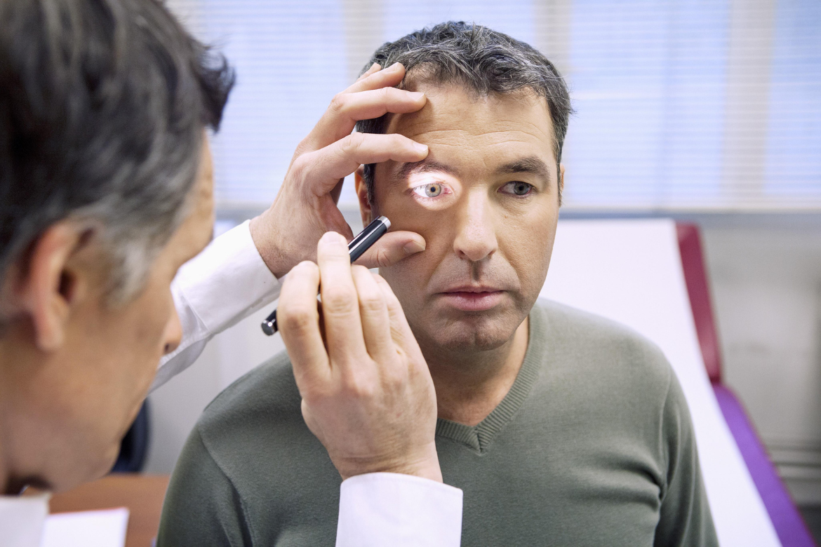
Diagnosis OF Retina Detachment:
The most common methods of diagnosing retinal detachment include
Optical Coherence Tomography: In the most common and simple method of diagnosis, your ophthalmologist will put drops in your eye to dilate and widen the pupil. Then, they will look through a special lens to check your retina for any damages or changes or damages, this is known as optical coherence tomography (OCT).
Treatments for Retina Detachment:
Treatment for retinal detachment usually involves surgery, and the types of procedures usually involved are as follows.
At the beginning of this procedure, your ophthalmologist will put a gas bubble inside your eye. This will push the retina into place so it can heal properly. Afterwards, your doctor will instruct you to keep your head in a very specific position for a few days. This position will keep the bubble in the right place. As your eye heals over time, your body makes fluid that fills the eye and this fluid will replace the gas bubble.
This procedure is done to remove the vitreous fluid that’s pulling on the retina. The vitreous fluid will be replaced with an air, gas, or oil bubble. The bubble will push the retina into place so it can heal properly. If an oil bubble is used, then it will be removed a few months later. If an air or gas bubble is used, you will not be allowed to fly in an aeroplane, travel to high altitudes, or scuba dive. This is because the change in altitude will cause the gas bubble to expand, thus increasing eye pressure.
In this procedure, a band of rubber or soft plastic is sewn to the outside of your eyeball. It will help in gently pressing your eye inward, thus causing the detached retina to heal against the eye wall. The scleral buckle cannot be seen in the eye and it is usually left on the eye permanently.
Prevention for Retina Detachment:
As many risks and factors contribute to retina detachment, it helps to know some precautionary measures that you can take beforehand. This will usually include,
- Regular eye care
- Regular appointments with your ophthalmologist to check your eye health if you have an eye condition.
- Stay safe and away from factors that affect the eyes or cause eye injuries.
- Watch out for warning signs such as ageing, diabetes, etc…