There are two main types of AMD: dry AMD, which accounts for about 80% of cases, and wet AMD, which requires more aggressive treatment. Common symptoms include blurry vision, distorted lines, and difficulty recognizing familiar faces. Risk factors include age over 50, smoking, and high blood pressure. Early diagnosis and treatments such as nutritional supplements and injections can help manage the condition effectively.
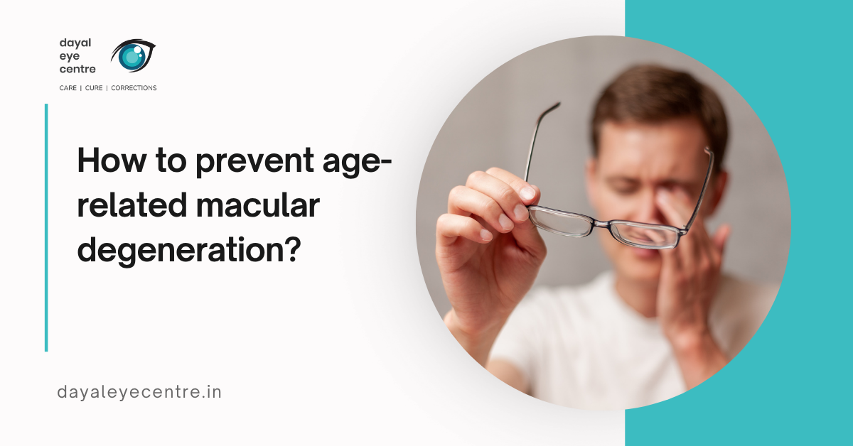
How AMD affects your vision over time
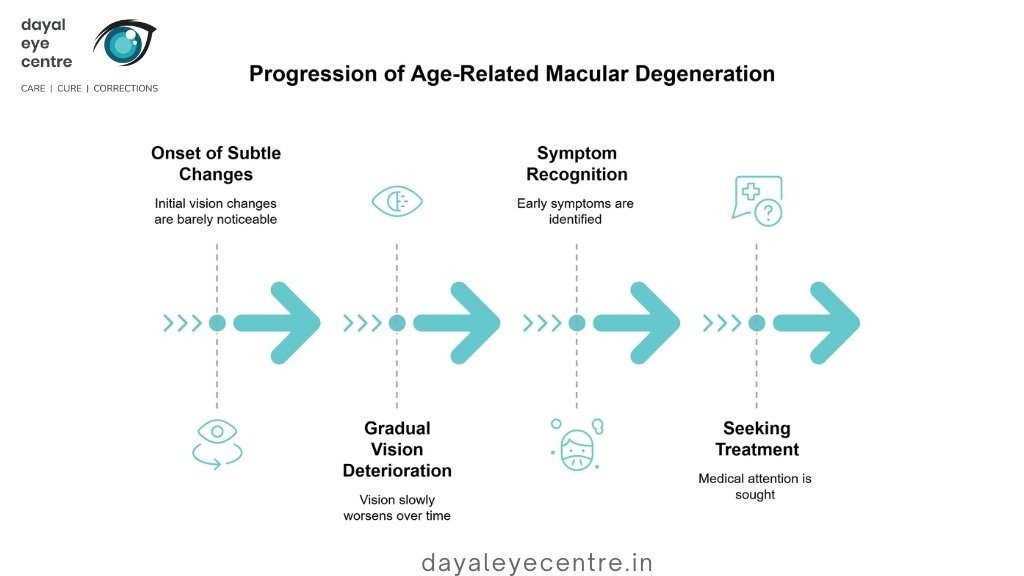
Age-related macular degeneration causes gradual changes in vision over time. These changes often begin so subtly that they may go unnoticed for years. Recognizing early symptoms is important, as timely evaluation and treatment can help slow disease progression and preserve vision.
Macular degeneration vision loss explained
Age-related macular degeneration (AMD) targets the macula, the central part of the retina responsible for sharp, detailed vision. The pattern of vision loss in AMD differs from that of many other eye conditions. Early signs may include wavy straight lines or colors appearing less vivid.
As the disease progresses, central vision becomes increasingly blurry. Blank or dark spots may develop, making everyday tasks more difficult. Imagine looking at a clock face—you may see the numbers around the edge clearly, but the hands in the center appear fuzzy.
Peripheral vs central vision in AMD
AMD has a distinctive feature: it affects only central vision. This area is essential for recognizing faces, reading, and performing detailed tasks. Peripheral (side) vision typically remains intact throughout the course of the disease.
People with AMD rarely become completely blind because their peripheral vision is preserved. Nevertheless, loss of central vision can significantly disrupt daily activities. Driving becomes difficult, reading is challenging, and recognizing faces is harder. Tasks requiring fine visual detail become increasingly difficult as the disease advances.
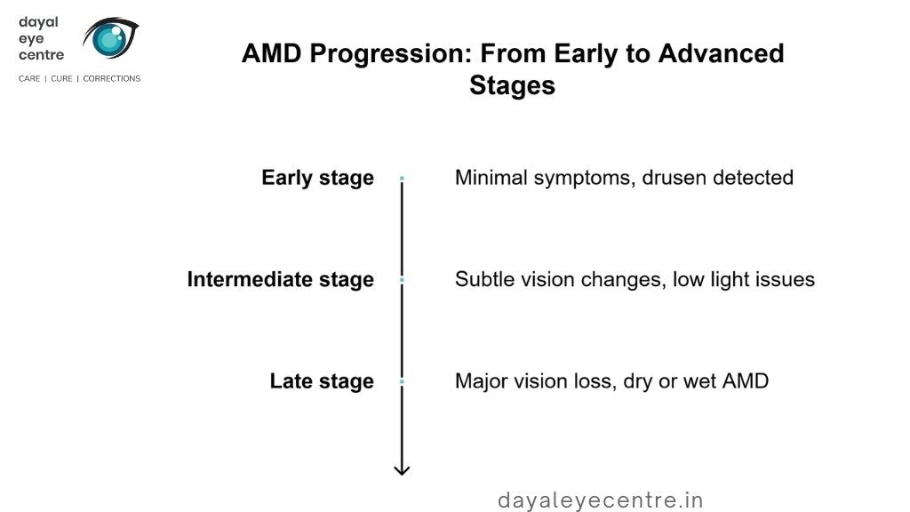
Progression from early to advanced stages
AMD progresses through defined stages, although not everyone experiences each stage:
- Early stage: Symptoms are minimal, but eye doctors may detect small yellow deposits called drusen. Vision usually remains normal at this stage.
- Intermediate stage: Subtle vision changes occur. You may have difficulty seeing in low light or distinguishing similar colors.
- Late (advanced) stage: Significant vision loss develops. Progression differs between types—dry AMD typically advances slowly over years, while wet AMD can cause rapid vision loss over weeks or months.
The speed of progression varies among individuals. In people with large drusen in both eyes, about 26% develop late-stage AMD within five years. The risk to the second eye increases when one eye already has advanced AMD, with incidence rates of 15.2 cases per 100 person-years for wet AMD and 11.2 cases per 100 person-years for dry AMD.
Identifying Early Signs and Symptoms of AMD
Early warning signs of age-related macular degeneration are important to recognize so doctors can intervene promptly. Subtle changes in the eyes may appear before noticeable vision loss occurs.
Visual distortions and blind spots
A key early sign of AMD is metamorphopsia—a visual distortion that causes straight lines to appear wavy, bent, or curved. This is significant, as it may indicate progression of the condition and requires prompt medical attention. People may also notice blurry areas or blind spots in their central vision. These can appear as dark, hazy areas or “whiteouts” in the center of vision and may enlarge over time.
Changes in reading and face recognition
Everyday activities often reveal early signs of macular degeneration. Reading becomes more difficult because words may appear blurry, even with appropriate glasses. Brighter lighting may be needed for reading or detailed tasks. Many people also experience difficulty recognizing faces, which can significantly affect social interactions. Studies show that even mild vision loss can impair facial recognition, sometimes leading to embarrassment, social withdrawal, and increased dependence on others. Individuals may rely on cues such as hairstyles or walking patterns, but these strategies are often unreliable.
When to see an eye doctor
You should see an eye doctor promptly if you notice any of the following:
- Straight lines appearing wavy or distorted
- Blurred central vision or blank spots
- Difficulty seeing fine details
- Increased difficulty reading or recognizing faces
- Worsening vision in low-light conditions
Early AMD often causes no obvious symptoms, making regular eye examinations essential, especially after the age of 60. AMD may affect one or both eyes, and changes can go unnoticed if only one eye is involved, as the unaffected eye may compensate. Early detection offers the best opportunity for effective management.
Getting Diagnosed: What to Expect During an Eye Exam
Accurate diagnosis is the foundation of effective macular degeneration treatment. A comprehensive eye examination is necessary to evaluate symptoms and confirm the condition.
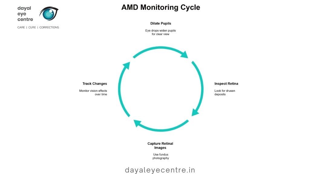
OCT and fluorescein angiography
Optical coherence tomography (OCT) is one of the most important advances in diagnostic imaging for macular degeneration. This non-invasive technique provides detailed cross-sectional images of the retina. OCT identifies areas where the retina has thinned, thickened, or swollen due to fluid accumulation—critical information for determining the most appropriate treatment approach.
Fluorescein angiography may be performed in patients suspected of having wet AMD. In this procedure, a special dye is injected into a vein in the arm and tracked as it flows through the retinal blood vessels. Areas of fluorescence can reveal leaking blood vessels characteristic of wet AMD. In some cases, indocyanine green angiography is added to help identify specific subtypes of macular degeneration.
Using the Amsler grid at home
The Amsler grid is a simple yet valuable tool for monitoring vision changes between doctor visits. This square grid with a central dot helps detect early visual distortions. To use it:
- Hold the grid approximately 12–14 inches from your eyes (normal reading distance).
- Cover one eye and focus on the center dot.
- Note any wavy, dim, distorted, or missing lines.
- Repeat the test with the other eye.
- Test weekly and contact your doctor immediately if you notice any changes.
Regular use of the Amsler grid can help detect early progression from dry to wet AMD, making it an important part of ongoing eye care.
Managing AMD: From Supplements to Injections
Although there is currently no cure for age-related macular degeneration, several treatments can slow disease progression and help preserve vision. Treatment options depend on whether AMD is dry or wet and the stage of the condition.
AREDS2 formula and dietary support
The Age-Related Eye Disease Study 2 (AREDS2) identified a specific vitamin and mineral supplement that reduces the risk of progression to advanced AMD by approximately 25% over five years. This formulation is most beneficial for individuals with:
- Intermediate AMD in one or both eyes
- Advanced AMD in one eye, to help protect the unaffected eye
The AREDS2 formula includes:
- 500 mg vitamin C
- 400 IU vitamin E
- 80 mg zinc (or 25 mg in the reduced formulation)
- 2 mg copper
- 10 mg lutein
- 2 mg zeaxanthin
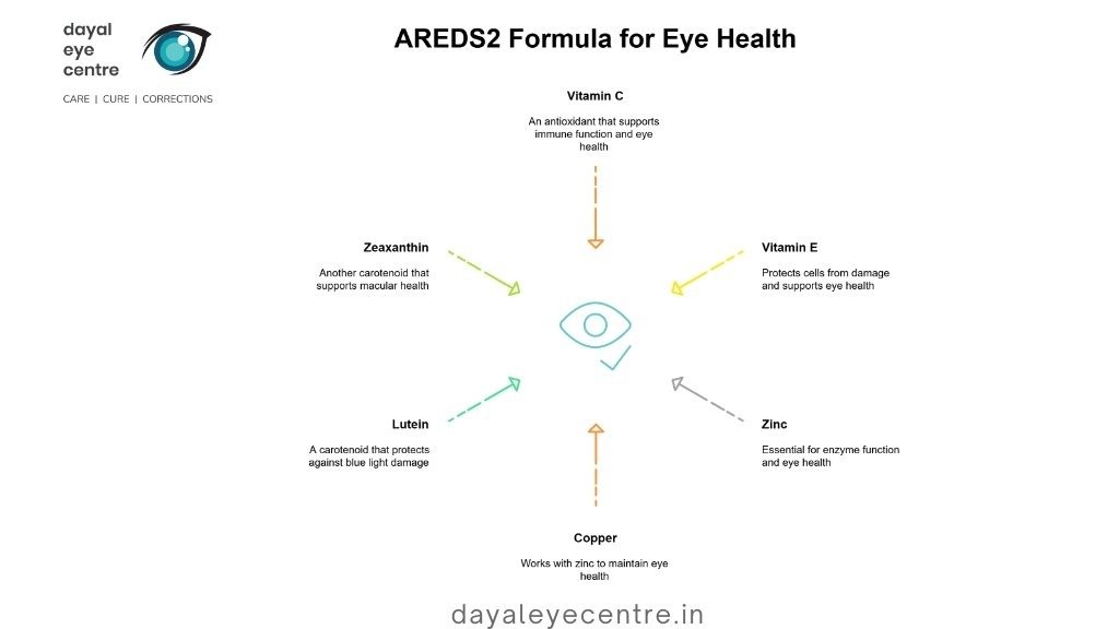
People with low dietary intake of lutein and zeaxanthin showed a 26% lower risk of developing advanced AMD when they took supplements. The Mediterranean diet has also demonstrated promising results, with a 25% reduction in the risk of disease progression.
Anti-VEGF therapy for wet AMD
Anti-VEGF medications are an effective way to control wet AMD. These drugs slow or stop abnormal blood vessel growth and leakage. The eye is numbed before the medication is administered through an injection.
Current anti-VEGF treatments include Avastin, Lucentis, Eylea, Beovu, Vabysmo, and Susvimo. Most patients require injections every 4–12 weeks. Newer options, such as Vabysmo, may allow treatment intervals of up to four months. Research shows that these therapies help about 90% of patients maintain their vision and improve vision in approximately one-third of cases..
Vision rehabilitation and assistive tools
Vision rehabilitation helps individuals maximize their remaining vision through specialized training and assistive devices. Options include:
- Non-optical devices: Large-print materials, audiobooks, tactile labels
- Electronic aids: Video magnifiers, OCR devices, smartphones with accessibility features
- Specialized eyewear: Telescopic lenses, magnifiers, and contrast-enhancing lenses
Rehabilitation specialists teach adaptive techniques that support independence despite vision changes and help identify solutions tailored to individual visual needs.
Emerging treatments and clinical trials
Research continues to advance in several promising areas:
- Gene therapy: Treatments such as RGX-314 and ADVM-022 aim to provide long-lasting anti-VEGF effects with a single treatment
- Stem cell therapy: Clinical trials are evaluating retinal pigment epithelium (RPE) cell replacement for dry AMD
- Complement inhibitors: New drugs targeting inflammatory pathways involved in geographic atrophy
- Port delivery systems: Implantable devices that release medication gradually and require refills only every six months
Ongoing clinical trials continue to expand treatment possibilities, offering hope for improved management of both dry and wet AMD.
Conclusion
Current understanding of age-related macular degeneration highlights its impact on millions of people worldwide. AMD presents a significant challenge for aging adults, primarily affecting central vision while usually preserving peripheral vision. As the disease progresses, patients often retain some visual function but experience increasing difficulty with reading and face recognition.
Early detection plays a critical role in treatment success. Adults over 50, particularly those with risk factors such as smoking or high blood pressure, should undergo regular eye examinations. These checkups serve as the first line of defense, while tools such as the Amsler grid help monitor vision changes between visits.
Several treatments can slow disease progression and preserve vision after diagnosis. The AREDS2 formula significantly benefits patients with intermediate or late-stage AMD, reducing progression by approximately 25% over five years. Anti-VEGF injections are highly effective for wet AMD, helping about 90% of patients maintain stable vision. Vision rehabilitation provides practical strategies and technologies to help individuals make the most of their remaining sight.
Ongoing research continues to bring hope. Advances in gene therapy, stem cell treatments, and drug delivery systems may lead to more effective treatment options. Although AMD currently has no cure, these developments point toward better outcomes for patients.
This guide aims to support a deeper understanding of AMD, whether you are living with the condition or supporting someone who is. Staying informed and acting promptly remain the most effective ways to protect vision and quality of life.
FAQs
What are the main types of age-related macular degeneration (AMD)?
There are two primary types of AMD: dry AMD, which accounts for about 80% of cases, and wet AMD. Dry AMD progresses slowly over years, while wet AMD can cause rapid vision deterioration within weeks or months.
How does AMD affect vision over time?
AMD primarily affects central vision, leading to blurriness, difficulty recognizing faces, and distortion of straight lines. Peripheral vision usually remains intact. As the disease progresses, blank spots may develop in central vision, making daily activities more challenging.
What are the early signs of AMD to watch for?
Early signs include difficulty reading or recognizing faces, needing brighter light for close-up tasks, and seeing straight lines as wavy or distorted. Blurry or dark spots may also appear in central vision.
How is AMD diagnosed?
AMD is typically diagnosed through a comprehensive eye examination, including pupil dilation, retinal imaging, and tests such as optical coherence tomography (OCT) and fluorescein angiography. The Amsler grid is also useful for monitoring vision changes at home.
What treatments are available for AMD?
Treatment depends on the type and stage of AMD. Dry AMD may benefit from the AREDS2 vitamin formula to slow progression, while wet AMD is commonly treated with anti-VEGF injections. Vision rehabilitation and assistive devices help manage vision loss, and emerging therapies such as gene and stem cell treatments are under active investigation.