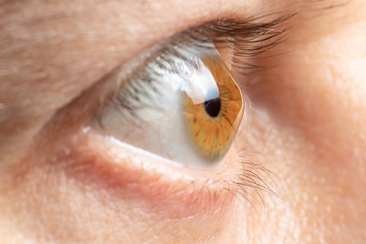
It refers to the condition in which your cornea -a transparent part of your eye that covers the iris and the pupil and is responsible for allowing light inside your eye and protecting it against infections- starts thinning and gradually bulges into a cone shape. A cone-shaped cornea causes blurred vision and may cause sensitivity to light and glare. Usually, it occurs in both eyes but in some cases, it might affect one eye more than the other.
Symptoms Of Keratoconus
- Blurred vision
- Increased sensitivity to bright light and glare
- Changing eye power with frequent changes in eyeglass prescriptions
- Sudden worsening or clouding of vision
- Double vision
Causes Of Keratoconus
- Not having enough protective antioxidants in your cornea. This can weaken your collagen and lead to an increase in harmful byproducts in the eye, thus bulging your cornea.
- A family history of Keratoconus.
- Rubbing your eyes vigorously
- Certain conditions like retinitis pigmentosa, Down syndrome, Ehlers-Danlos syndrome, hay fever, and asthma also increase your chances of being affected with Keratoconus.
- Inflammation from eye allergies, asthma, or atopic eye disease can break down the tissue of the cornea
Diagnosis
Eye Refraction: In this test, our eye doctor uses special equipment to measure your eyes to check for vision problems. Our ophthalmologist might ask you to look through a device that contains wheels of different lenses, known as the phoropter to accurately judge which combination gives you the sharpest vision. Some doctors may use a hand-held instrument such as the retinoscope to evaluate your eyes.
Slit-Lamp Examination: In this test, a vertical beam of light directs into your eye and then through a low-powered microscope, he might asses your eye. To diagnose keratoconus, they might evaluate the shape of your cornea and looks for other potential problems and conditions in your eye.
Keratometry: In this test, your eye doctor focuses a circle of light on your cornea to measure the reflection and examine the basic shape of your cornea.
Computerized Corneal Mapping: These include specialized photographic tests, such as corneal tomography and corneal topography, which are used to record images to create a detailed shape map of your cornea. Corneal topography can also measure the thickness of your cornea and can often detect early signs of keratoconus before the disease is externally visible.
Treatment For Keratoconus
- Slowing the rate of progression
- Improving your vision
These are usually prescribed for a very advanced stage of Keratoconus when your cornea starts morphing into a very irregular shape. Instead of touching the cornea like regular lenses, scleral lenses sit on the white part of your eye (sclera) and hover over your cornea.Surgery is usually done if you have corneal scarring, extreme thinning, or poor vision, if the lenses or eyeglasses are not working for you, or if you can’t wear prescribed lenses.
Living with Keratoconus
- Avoid rubbing your eyes
- Wear sun protection for your eyes
- Avoid eye allergies
- Eat a healthy and balanced diet
- Optimising your contact lens to the maximum.